Structural analysis of 2qgn: Difference between revisions
Choojinhsien (talk | contribs) |
Choojinhsien (talk | contribs) |
||
| (119 intermediate revisions by the same user not shown) | |||
| Line 1: | Line 1: | ||
==General Properties of 2qgn== | |||
General information collected from PDB indicates that : | |||
(a) 2qgn is a tRNA isopentenyl transferase isolated from Bacillus holodurans c-125, expressed in Escherichia Coli. | |||
(b) Also known as tRNA delta(2)-isopentenylpyrophosphate transferase, belonging to the IPP transferase family | |||
(c) Resolution of 2.4 angstroms, with an r-value of 0.225. | |||
(d) Ligand chemical component identified as sulfate ion. | |||
==Protein Sequence in FASTA format== | ==Protein Sequence in FASTA format== | ||
>gi|152149497|pdb|2QGN|A Chain A, Crystal Structure Of Trna Isopentenylpyrophosphate Transferase (Bh2366) From Bacillus Halodurans, Northeast Structural Genomics Consortium Target Bhr41. | >gi|152149497|pdb|2QGN|A Chain A, Crystal Structure Of Trna Isopentenylpyrophosphate Transferase (Bh2366) From Bacillus Halodurans, Northeast Structural Genomics Consortium Target Bhr41. | ||
| Line 8: | Line 20: | ||
==Structure of Protein== | ==Structure of Protein== | ||
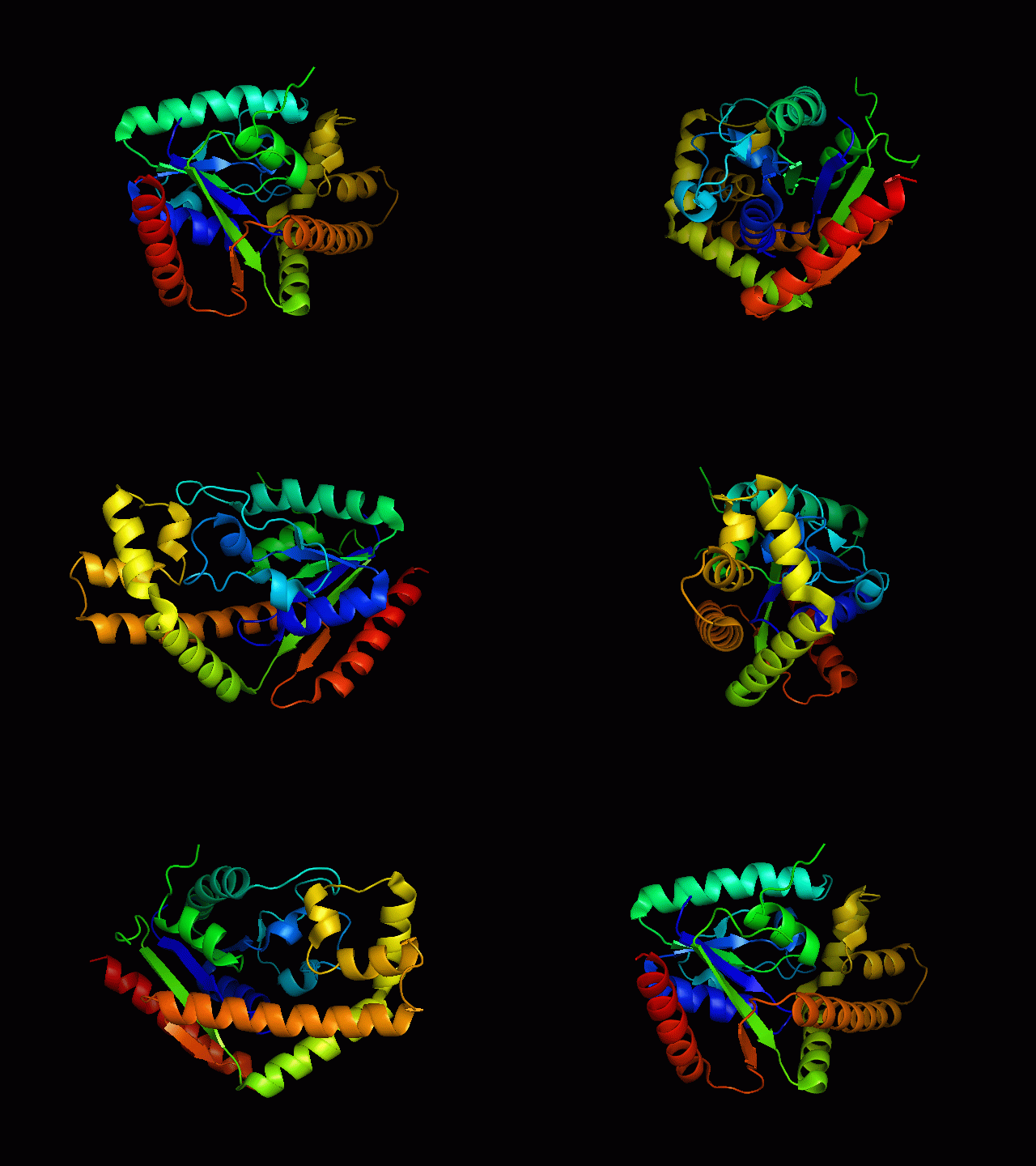
[[Image: | [[Image:2qgnA3.png|framed|'''Figure 1'''<BR>Structure of tRNA isopentenyl transferase 1 showing helix, sheet and loop. Image constructed from PyMOL.|none]]<BR> | ||
== | ==Secondary Structure and Location of P-loop (ATP binding site)== | ||
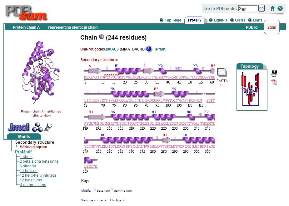
Analysis of the secondary structure acquired from Protein Data Bank showed results as displayed below : | Analysis of the secondary structure acquired from Protein Data Bank showed results as displayed below : | ||
[[Image:Secondary.jpg|framed|none]] | [[Image:Secondary.jpg|framed|none]] | ||
==Surface | P-loop region in the protein was found via a conserved GxTxxGK(T/S) motif, similar to the P-loop in kinases as well as other nucleotide binding proteins. From the secondary structure, the dots highlighted in red proposes the possible location of P-loop. | ||
[[Image: | |||
==Surface Structure== | |||
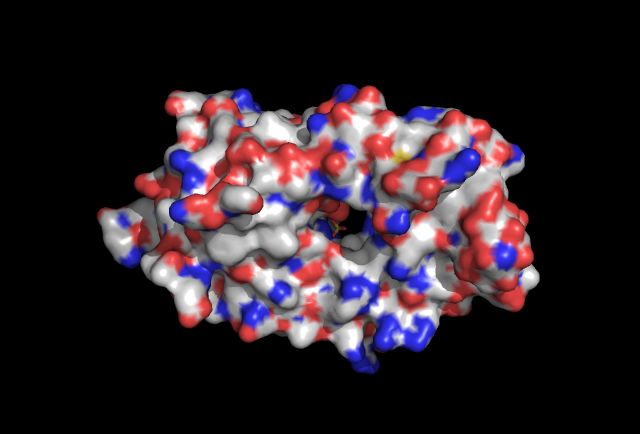
[[Image:surface view.jpg|framed|'''Figure 1.1''' Surface view of protein with ligand found in the cavitiy of the protein.|none]] | |||
==Electrostatic Surface Potential== | |||
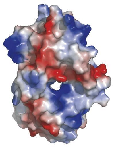
[[Image:esp.jpg|framed|'''Figure 1.2''' Electrostatic surface potential of 2qgn. Positve regions indicated by blue, negatively charged in red.|none]] | |||
==Domains== | |||
2qgnA is composed of two main domains. CATH analysis of 2qgn resulted in the finding of two main domains composing 2qgnA. | |||
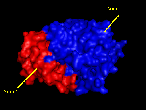
Domain 1 ranges from residue 2-200 and residue 283-314. Domain 2 encompasses residues stretching from 201-282. Topology of Domain 1 is known to be of Rossmann A-B-A fold, and is a representative of the homologous superfamily of P-loop containing nucleotide triphosphate hydrolases. Domain 2 is yet to be classified. | |||
[[Image:domain pic3.jpg|framed|'''Figure 4''' <BR>Two main domains exhibited by tRNA isopentenyl transferase(2qgnA). Blue regions denote first domain while Red regions underlies second domain.|none]]<BR> | |||
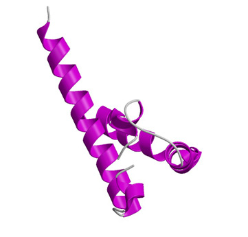
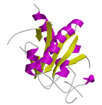
[[Image:domain--1.jpg|framed|left|'''Figure 5'''Ribbon structure of domain 2 signified by red regions in Figure 4.]][[Image:domain--2.jpg|framed|left|'''Figure 6'''Ribbon structure of domain 1 denoted by blue regions in Figure 4.]]<BR> | |||
<BR><BR><BR><BR><BR><BR><BR><BR><BR><BR><BR><BR><BR><BR><BR><BR><BR><BR><BR><BR><BR><BR> | |||
==Surface Topography== | |||
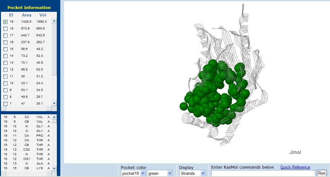
[[Image:pocket.jpg|framed|'''Figure 3''' Putative pocket in tRNA isopentenyl transferase. A total of 19 pockets were found in 2qgnA, displayed in green is the pocket with the largest area and volume.|none]] | |||
==Ligand Binding Sites and Surface Clefts== | ==Ligand Binding Sites and Surface Clefts== | ||
[[Image:ligand.png|framed|none]] | [[Image:ligand.png|framed|none]] | ||
LIGPLOT of protein shows protein-ligand interaction. The interactions shown are those mediated by hydrogen bonds and by | |||
hydrophobic contacts. Hydrogen bonds indicated by dashed lines between the atoms involved, while hydrophobic contacts are | |||
represented by an arc with spokes radiating towards the ligand atoms they contact. The contacted atoms are shown with spokes | |||
radiating back. | |||
[[Image:surface cleft.png|framed|none]] | [[Image:surface cleft.png|framed|none]] | ||
==Protein-ligand interaction == | |||
'''Hydrophillic binding sites''' | |||
[[Image:hydrophillic.jpg|framed|none]] | |||
'''Bridged-H-bond binding sites''' | |||
[[Image:H-bond.jpg|framed|none]] | |||
'''Hydrophobic binding sites''' | |||
[[Image:hydrophobic.jpg|framed|none]] | |||
==Conserved residues from Clustal alignment== | |||
Multiple sequence alignment from ClustalX allowed conserved regions in 2qgn and related species to be found. | |||
[[Image:conserved regions.jpg|framed|'''Figure 5''' Conserved regions among various species were shown in red, with their respective residues labelled. Yellow sphere shows the location of the ligand. Image was constructed from PyMOL. |none]]<BR> | |||
== Structural Alignment== | == Structural Alignment== | ||
'''Dali Output''' | |||
PDB entry code for 2qgn was loaded onto DALI server to search for structurally similar neighbours. Displayed below are the results from DALI search :- | |||
[[Image:Dali output3.jpg|framed|none]] | |||
DALI output describes the following : | |||
''Z score'' , the statistical significance of the similarity between protein-of-interest and other neighbourhood protines. The program optimises a weighted sum of similarities of intramolecular distances. | |||
''Root Mean Square Distance (RMSD)'', root-mean-square deviation of C-alpha atoms in the least-squares superimposition of the structurally equivalent C-alpha atoms. As in indicated in DALI, rmsd is not optimised and is only reported for information. | |||
''lali'', the number of structurally equivalent residues. | |||
''nres'', or the total number of amino acids in the hit protein. | |||
''%id'' - percentage of identical amino acids over structurally equivalent residues. | |||
A total of 527 hits were found from DALI search, nonetheless only the first 20 hits that may be of significance were shown on the figure. | |||
'''Profunc''' | |||
''Related protein sequences'' | |||
[[Image:profunc2.jpg|framed|none]] | |||
''Proteins with similar fold retrived from SSM (Secondary Structure Matching)'' | |||
[[Image:ssm2.jpg|framed|none]] | |||
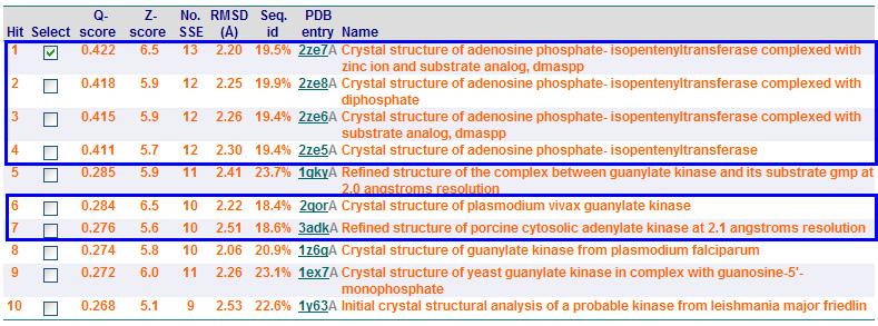
From Profunc, similarities of related proteins and proteins with similar fold to query protein were compared with results from DALI. 2qgnA is the query protein highlighted in black in all tables. 2crm, 2crr and 2crq were both found in DALI and Profunc(highlighted in red). On the other hand, 2ze5,2ze6,2ze7 and 2ze8, as well as 3adk and 2qor were also found in DALI output(highlighted in blue). | |||
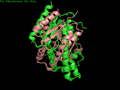
Based on the outcome of DALI and Profunc, PDB files of each structurally similar protein was obtained from PDB. These were each superimposed against 2qgn using the PyMOL software, to compare the structural similiarity. Results are as below : | |||
PDB | |||
[[Image: | [[Image:2qgn421.jpg|framed|left|'''Figure 7''' 3crq superimposed against 2qgn via PyMOL. 2qgn indicated in green.]][[Image:2qgn2.png|framed|left|'''Figure 8''' 3crm superimposed against 2qgn via PyMOL. 2qgn indicated in green.]]<BR> <BR><BR><BR><BR><BR><BR><BR><BR><BR><BR><BR><BR><BR><BR><BR><BR><BR><BR><BR><BR><BR><BR> | ||
[[Image:2qgn.png|framed|left|'''Figure 9''' 2ze7 superimposed against 2qgn via PyMOL. 2qgn indicated in green.]][[Image:2qor.jpg|framed|left|'''Figure 10''' 2qor superimposed against 2qgn via PyMOL. 2qgn indicated in green]]<BR> | |||
<BR><BR><BR><BR><BR><BR><BR><BR><BR><BR><BR><BR><BR><BR><BR><BR><BR><BR><BR><BR><BR><BR> | |||
As indicated by the figures above, each structures were structurally similar to 2qgn, suggesting that they could have functionally similar properties. Nonetheless,notice that 2-qor is only partially similar to 2qgn structure. | |||
As the Z-score decreases for the DALI output, the structural similarity decreases as well. For this reason, functional analysis of 2qgn was only done for DALI outputs with lali scores higher than 200. | |||
Latest revision as of 06:37, 10 June 2008
General Properties of 2qgn
General information collected from PDB indicates that :
(a) 2qgn is a tRNA isopentenyl transferase isolated from Bacillus holodurans c-125, expressed in Escherichia Coli.
(b) Also known as tRNA delta(2)-isopentenylpyrophosphate transferase, belonging to the IPP transferase family
(c) Resolution of 2.4 angstroms, with an r-value of 0.225.
(d) Ligand chemical component identified as sulfate ion.
Protein Sequence in FASTA format
>gi|152149497|pdb|2QGN|A Chain A, Crystal Structure Of Trna Isopentenylpyrophosphate Transferase (Bh2366) From Bacillus Halodurans, Northeast Structural Genomics Consortium Target Bhr41. XKEKLVAIVGPTAVGKTKTSVXLAKRLNGEVISGDSXQVYRGXDIGTAKITAEEXDGVPHHLIDIKDPSE SFSVADFQDLATPLITEIHERGRLPFLVGGTGLYVNAVIHQFNLGDIRADEDYRHELEAFVNSYGVQALH DKLSKIDPKAAAAIHPNNYRRVIRALEIIKLTGKTVTEQARHEEETPSPYNLVXIGLTXERDVLYDRINR RVDQXVEEGLIDEAKKLYDRGIRDCQSVQAIGYKEXYDYLDGNVTLEEAIDTLKRNSRRYAKRQLTWFRN KANVTWFDXTDVDFDKKIXEIHNFIAGKLEEKSKLEHHHHHH
Structure of Protein
Secondary Structure and Location of P-loop (ATP binding site)
Analysis of the secondary structure acquired from Protein Data Bank showed results as displayed below :
P-loop region in the protein was found via a conserved GxTxxGK(T/S) motif, similar to the P-loop in kinases as well as other nucleotide binding proteins. From the secondary structure, the dots highlighted in red proposes the possible location of P-loop.
Surface Structure
Electrostatic Surface Potential
Domains
2qgnA is composed of two main domains. CATH analysis of 2qgn resulted in the finding of two main domains composing 2qgnA.
Domain 1 ranges from residue 2-200 and residue 283-314. Domain 2 encompasses residues stretching from 201-282. Topology of Domain 1 is known to be of Rossmann A-B-A fold, and is a representative of the homologous superfamily of P-loop containing nucleotide triphosphate hydrolases. Domain 2 is yet to be classified.
Surface Topography
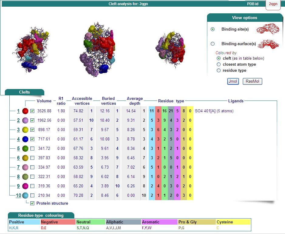
Ligand Binding Sites and Surface Clefts
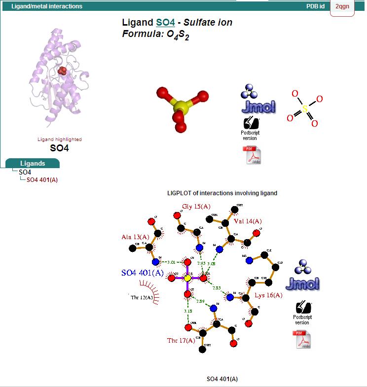
LIGPLOT of protein shows protein-ligand interaction. The interactions shown are those mediated by hydrogen bonds and by
hydrophobic contacts. Hydrogen bonds indicated by dashed lines between the atoms involved, while hydrophobic contacts are
represented by an arc with spokes radiating towards the ligand atoms they contact. The contacted atoms are shown with spokes
radiating back.
Protein-ligand interaction
Hydrophillic binding sites
Bridged-H-bond binding sites
Hydrophobic binding sites
Conserved residues from Clustal alignment
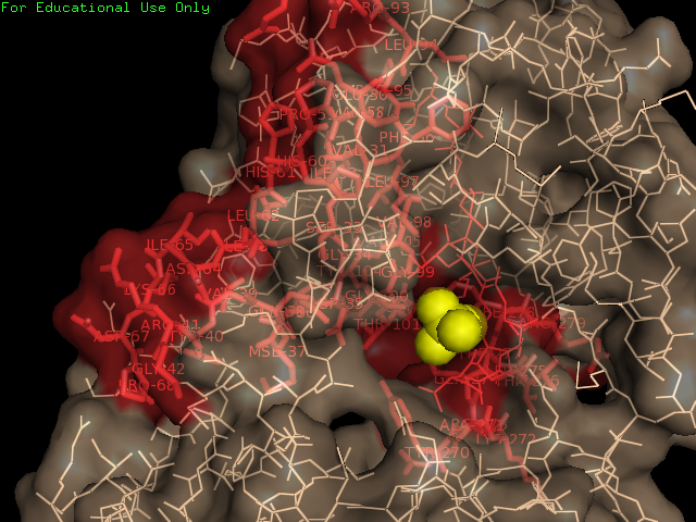
Multiple sequence alignment from ClustalX allowed conserved regions in 2qgn and related species to be found.
Structural Alignment
Dali Output
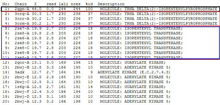
PDB entry code for 2qgn was loaded onto DALI server to search for structurally similar neighbours. Displayed below are the results from DALI search :-
DALI output describes the following :
Z score , the statistical significance of the similarity between protein-of-interest and other neighbourhood protines. The program optimises a weighted sum of similarities of intramolecular distances.
Root Mean Square Distance (RMSD), root-mean-square deviation of C-alpha atoms in the least-squares superimposition of the structurally equivalent C-alpha atoms. As in indicated in DALI, rmsd is not optimised and is only reported for information.
lali, the number of structurally equivalent residues.
nres, or the total number of amino acids in the hit protein.
%id - percentage of identical amino acids over structurally equivalent residues.
A total of 527 hits were found from DALI search, nonetheless only the first 20 hits that may be of significance were shown on the figure.
Profunc
Related protein sequences
Proteins with similar fold retrived from SSM (Secondary Structure Matching)
From Profunc, similarities of related proteins and proteins with similar fold to query protein were compared with results from DALI. 2qgnA is the query protein highlighted in black in all tables. 2crm, 2crr and 2crq were both found in DALI and Profunc(highlighted in red). On the other hand, 2ze5,2ze6,2ze7 and 2ze8, as well as 3adk and 2qor were also found in DALI output(highlighted in blue).
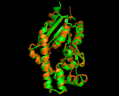
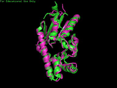
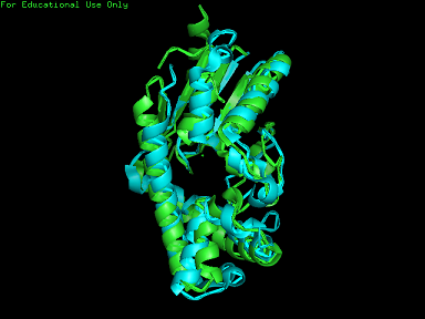
Based on the outcome of DALI and Profunc, PDB files of each structurally similar protein was obtained from PDB. These were each superimposed against 2qgn using the PyMOL software, to compare the structural similiarity. Results are as below :
As indicated by the figures above, each structures were structurally similar to 2qgn, suggesting that they could have functionally similar properties. Nonetheless,notice that 2-qor is only partially similar to 2qgn structure.
As the Z-score decreases for the DALI output, the structural similarity decreases as well. For this reason, functional analysis of 2qgn was only done for DALI outputs with lali scores higher than 200.
