Results (1zkd): Difference between revisions
No edit summary |
No edit summary |
||
| Line 1: | Line 1: | ||
==Structure of Hypothetical Protein LOC55471 Isoform 1== | ==Structure of Hypothetical Protein LOC55471 Isoform 1== | ||
===Comparing Structure of Proteins=== | |||
Dali shows a few proteins with similar structure to 1zkd. They are 2ex4 and 1im8 which shows the highest Z-value of 11.7 and 11.6 respectively. 2ex4 is a human methyltransferase with S-adenosylhomocysteine and 1im8 is found to be a methyltransferase with a bound S-adenosylhomocysteine from the crystal structure of YecO from Haemophilus influenzae (HI0319). | Dali shows a few proteins with similar structure to 1zkd. They are 2ex4 and 1im8 which shows the highest Z-value of 11.7 and 11.6 respectively. 2ex4 is a human methyltransferase with S-adenosylhomocysteine and 1im8 is found to be a methyltransferase with a bound S-adenosylhomocysteine from the crystal structure of YecO from Haemophilus influenzae (HI0319). | ||
| Line 76: | Line 78: | ||
RMSD min – max: 2.3A | RMSD min – max: 2.3A | ||
===Ligand and Binding Sites=== | |||
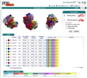
[[Image:Binding sites.JPG|thumb|Binding Sites]] | |||
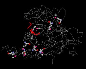
[[Image:Ligand.JPG|thumb|Conserved regions]] | |||
Revision as of 06:52, 6 June 2007
Structure of Hypothetical Protein LOC55471 Isoform 1
Comparing Structure of Proteins
Dali shows a few proteins with similar structure to 1zkd. They are 2ex4 and 1im8 which shows the highest Z-value of 11.7 and 11.6 respectively. 2ex4 is a human methyltransferase with S-adenosylhomocysteine and 1im8 is found to be a methyltransferase with a bound S-adenosylhomocysteine from the crystal structure of YecO from Haemophilus influenzae (HI0319).
SUMMARY: PDB/chain identifiers and structural alignment statistics
NR. STRID1 STRID2 Z RMSD LALI LSEQ2 %IDE REVERS PERMUT NFRAG TOPO PROTEIN
1:  3027-A 1zkd-A 56.8  0.0  349   349  100      0      0     1 S    STRUCTURAL GENOMICS, UNKNOWN FUNCTION    duf185  (rhodops
2:  3027-A 2ex4-A 11.7  3.0  185   221   12      0      0    22 S    TRANSFERASE      adrenal gland protein ad-003    (homo sapien
3:  3027-A 1im8-A 11.6  3.2  178   225   10      0      0    18 S    TRANSFERASE     yeco (methyltransferase, hypothetical pro
4:  3027-A 2gb4-A 10.8  3.3  184   231   13      0      0    19 S    TRANSFERASE      thiopurine s-methyltransferase (thiopurine
5:  3027-A 2fk7-A 10.7  3.8  186   277   14      0      0    19 S    TRANSFERASE      methoxy mycolic acid synthase 4         (mycobact
6:  3027-A 2ob1-A 10.2  3.9  196   319    9      0      0    23 S    TRANSFERASE      leucine carboxyl methyltransferase 1 (prot
7:  3027-A 2f8l-A 10.0  3.7  182   318    8      0      0    22 S    STRUCTURAL GENOMICS, UNKNOWN FUNCTION    hypothetical pro
8:  3027-A 2avn-A 10.0  3.7  183   242   11      0      0    20 S    STRUCTURAL GENOMICS, UNKNOWN FUNCTION    ubiquinoneMENAQU
9:  3027-A 2bzg-A  9.9  3.4  182   226   13      0      0    20 S    TRANSFERASE      thiopurine s-methyltransferase (thiopurine
10: 3027-A 2aot-A  9.8  4.3  182   285   13      0      0    23 S    TRANSFERASE      histamine n-methyltransferase (hmt)     (homo
1zkd is an unknown protein, and by using proteins similar to it, the functions of this unknown protein can be predicted. With 2ex4 and 1im8 showed by Dali to be the most similar, other tools are used to determine the similarity. Combinatorial Extension Method is used. Below shows the sequence alignment and structure alignment of the unknown protein with the proteins obtained from dali:
Alignment with 2ex4
1ZKD:A   24/25    WRYXELCLGHPEHGYYV--TRDPLGREGDFTTSPEISQXFGELLGLWSASVWKAAD-EPQ
2EX4:A   24/7     IEDEKQFYS----KAKTYWKQIPPTVDGMLGGYGHISSIDINSSRKFLQRFLREGPNKTG
1ZKD:A   81/82    TLRLIEIGPGRGTXXADALRALRVLPILYQSLSVHLVEINPVLRQKQQTLLAGI-RNIHW
2EX4:A   80/64    TSCALDCGAGIGRITKRLLLPL--------FREVDMVDITEDFLVQAKTYLGEEGKRVRN
1ZKD:A  140/141   HD-----SFEDVPEGPAVILANEYFDVLPIHQAIKRETGWHERVIEIGASGELVFGVAAD
2EX4:A  132/116   YFCCGLQDFTPEPDSYDVIWIQWVIGHLT-------------------------------
1ZKD:A  195/196   PIPGFEALLPPLARLSPPGAVFEWRP--DTEILKIASRVRDQGGAALIIDYG--HLRSDV
2EX4:A  161/145   ------------------------DQHLAEFLRRCKGSL-RPNGIIVIKDNMAQE-----
1ZKD:A  251/252   GDTFQAIASHSYADPLQHPGRADLTAHV---DFDALGRAAESIGARAHGPVTQG
2EX4:A  191/175   GVILDD---------------VDSSVCRDLDVVRRIICSAG---LSLLAEERQE
Alignment with 1im8
1ZKD:A   59/60    QXFGELLGLWSASVWKAADEPQTLRLIEIGPGRGTXXADALRALRVLPILYQSLSVHLVE
1IM8:A   42/40    SNIITAIGXLAERFV-----TADSNVYDLGCSRGAATLSARRNI-----NQPNVKIIGID
1ZKD:A  119/120   INPVLRQKQQTLLAGI---RNIHWHD--SFEDVPEGPAVILANEYFDVLPIHQAIKRETG
1IM8:A   92/90    NSQPXVERCRQHIAAYHSEIPVEILCNDIRHVEIKNASXVILNFTLQFLP----------
1ZKD:A  174/175   WHERVIEIGASGELVFGVAADPIPGFEALLPPLARLSPPGAVFEWRP--DTEILKIASRV
1IM8:A  142/140   ---------------------------------------------PEDRIALLTKIYEGL
1ZKD:A  232/233   RDQG--GAALIIDYG
1IM8:A  157/155   ---NPNGVLVLSEKF
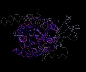
Structure Alignment with 2ex4 (Blue:1zkd, Purple: 2ex4)
Alignment Length: 294
Gaps (average per molecule): 53.5
Sequence Identity: 14.4%
RMSD min – max: 3.03A
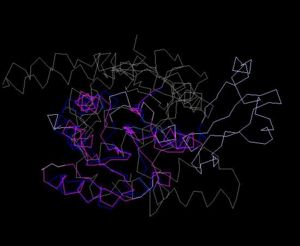
Structure Alignment with 1im8 (Blue:1zkd, Purple: 1im8)
Alignment Length: 195
Gaps (average per molecule): 38.5
Sequence Identity: 11%
RMSD min – max: 2.3A
Ligand and Binding Sites
Return to Report on 1zkd


