Results (1zkd): Difference between revisions
| Line 307: | Line 307: | ||
=== Evolutionary Tree === | === Evolutionary Tree === | ||
[[Image:Tree wit bootstrap.JPG|thumb| | [[Image:Tree wit bootstrap.JPG|thumb|700px]] | ||
Revision as of 06:46, 9 June 2007
Structure of Hypothetical Protein LOC55471 Isoform 1
Comparing Structure of Proteins
Dali shows a few proteins with similar structure to 1zkd. They are 2ex4 and 1im8 which shows the highest Z-value of 11.7 and 11.6 respectively. The higher the Z-value the more significant is the results. However, they are only 10-12% identical to the query protein. Nevertheless, these 2 proteins are used to compare with the query protein as these 10-12% identity may be at the binding site or ligand which will determine the functions. 2ex4 is a human methyltransferase with S-adenosylhomocysteine and 1im8 is found to be a methyltransferase with a bound S-adenosylhomocysteine from the crystal structure of YecO from Haemophilus influenzae (HI0319).
SUMMARY: PDB/chain identifiers and structural alignment statistics
NR. STRID1 STRID2  Z   RMSD LALI LSEQ2 %IDE REVERS PERMUT NFRAG TOPO PROTEIN
1:  3027-A 1zkd-A 56.8  0.0  349   349  100      0      0     1 S    STRUCTURAL GENOMICS, UNKNOWN FUNCTION    duf185  (rhodops
2:  3027-A 2ex4-A 11.7  3.0  185   221   12      0      0    22 S    TRANSFERASE      adrenal gland protein ad-003    (homo sapien
3:  3027-A 1im8-A 11.6  3.2  178   225   10      0      0    18 S    TRANSFERASE     yeco (methyltransferase, hypothetical pro 4:  3027-A 2gb4-A 10.8  3.3  184   231   13      0      0    19 S    TRANSFERASE      thiopurine s-methyltransferase (thiopurine
5:  3027-A 2fk7-A 10.7  3.8  186   277   14      0      0    19 S    TRANSFERASE      methoxy mycolic acid synthase 4         (mycobact
6:  3027-A 2ob1-A 10.2  3.9  196   319    9      0      0    23 S    TRANSFERASE      leucine carboxyl methyltransferase 1 (prot
7:  3027-A 2f8l-A 10.0  3.7  182   318    8      0      0    22 S    STRUCTURAL GENOMICS, UNKNOWN FUNCTION    hypothetical pro
8:  3027-A 2avn-A 10.0  3.7  183   242   11      0      0    20 S    STRUCTURAL GENOMICS, UNKNOWN FUNCTION    ubiquinoneMENAQU
9:  3027-A 2bzg-A  9.9  3.4  182   226   13      0      0    20 S    TRANSFERASE      thiopurine s-methyltransferase (thiopurine
10: 3027-A 2aot-A  9.8  4.3  182   285   13      0      0    23 S    TRANSFERASE      histamine n-methyltransferase (hmt)     (homo
1zkd is an unknown protein, and by using proteins similar to it, the functions of this unknown protein can be predicted. With 2ex4 and 1im8 showed by Dali to be the most similar, other tools are used to determine the similarity. Combinatorial Extension Method is used. Below shows the sequence alignment and structure alignment of the unknown protein with the proteins obtained from dali:
Alignment with 2ex4
1ZKD:A   24/25    WRYXELCLGHPEHGYYV--TRDPLGREGDFTTSPEISQXFGELLGLWSASVWKAAD-EPQ
2EX4:A   24/7     IEDEKQFYS----KAKTYWKQIPPTVDGMLGGYGHISSIDINSSRKFLQRFLREGPNKTG
1ZKD:A   81/82    TLRLIEIGPGRGTXXADALRALRVLPILYQSLSVHLVEINPVLRQKQQTLLAGI-RNIHW
2EX4:A   80/64    TSCALDCGAGIGRITKRLLLPL--------FREVDMVDITEDFLVQAKTYLGEEGKRVRN
1ZKD:A  140/141   HD-----SFEDVPEGPAVILANEYFDVLPIHQAIKRETGWHERVIEIGASGELVFGVAAD
2EX4:A  132/116   YFCCGLQDFTPEPDSYDVIWIQWVIGHLT-------------------------------
1ZKD:A  195/196   PIPGFEALLPPLARLSPPGAVFEWRP--DTEILKIASRVRDQGGAALIIDYG--HLRSDV
2EX4:A  161/145   ------------------------DQHLAEFLRRCKGSL-RPNGIIVIKDNMAQE-----
1ZKD:A  251/252   GDTFQAIASHSYADPLQHPGRADLTAHV---DFDALGRAAESIGARAHGPVTQG
2EX4:A  191/175   GVILDD---------------VDSSVCRDLDVVRRIICSAG---LSLLAEERQE
Alignment with 1im8
1ZKD:A   59/60    QXFGELLGLWSASVWKAADEPQTLRLIEIGPGRGTXXADALRALRVLPILYQSLSVHLVE
1IM8:A   42/40    SNIITAIGXLAERFV-----TADSNVYDLGCSRGAATLSARRNI-----NQPNVKIIGID
1ZKD:A  119/120   INPVLRQKQQTLLAGI---RNIHWHD--SFEDVPEGPAVILANEYFDVLPIHQAIKRETG
1IM8:A   92/90    NSQPXVERCRQHIAAYHSEIPVEILCNDIRHVEIKNASXVILNFTLQFLP----------
1ZKD:A  174/175   WHERVIEIGASGELVFGVAADPIPGFEALLPPLARLSPPGAVFEWRP--DTEILKIASRV
1IM8:A  142/140   ---------------------------------------------PEDRIALLTKIYEGL
1ZKD:A  232/233   RDQG--GAALIIDYG
1IM8:A  157/155   ---NPNGVLVLSEKF
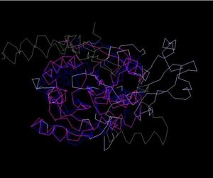
Structure Alignment with 2ex4 (Blue:1zkd, Purple: 2ex4)
Alignment Length: 294
Gaps (average per molecule): 53.5
Sequence Identity: 14.4%
RMSD min – max: 3.03A
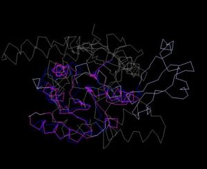
Structure Alignment with 1im8 (Blue:1zkd, Purple: 1im8)
Alignment Length: 195
Gaps (average per molecule): 38.5
Sequence Identity: 11%
RMSD min – max: 2.3A
Ligand and Binding Sites
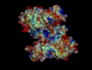
Surface Properties
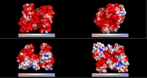
Red shows negatively charge and blue shows positively charge
Domain
Domain Identification Using Pfam
DUF185: domain 1 of 1, from 64 to 299: score 227.1, E = 3.9e-65
                  *->alArwllveykllgyPYadlnlvElGaGrGtaielmsdlLryiarlv
                     +l++w + ++k+ ++P   l+l E+G+GrGt   +m+d+Lr+  r+
      query    64    LLGLWSASVWKAADEP-QTLRLIEIGPGRGT---MMADALRA-LRVL 105
                  PdvyartryylvEiSprLaarQketLapkvaplGhdskveieatdlsglv
                  P +y+ ++++lvEi+p L+++Q++ La                 ++  ++
      query   106 PILYQSLSVHLVEINPVLRQKQQTLLA-----------------GIR-NI 137
                  rWhdasileedPdgvptvliaNEVlDalPHDlvrfdkrgggwyErhVlvd
                   Whd s +e++P+g p v++aNE +D lP  +++ +kr+ gw+Er V ++
      query   138 HWHD-S-FEDVPEG-PAVILANEYFDVLP--IHQAIKRETGWHER-V-IE 180
                  ldgdfrlvysqeldplaglaltlreaaldPVKstkklvpsalskllpkll
                    + ++lv+++++dp  g+                            ++l
      query   181 IGASGELVFGVAADPIPGFEAL------------------------LPPL 206
                  ppaeevgygtEvYsParllellqalaerLpahrGrlLaiDYGhlaseyyh
                   +   +g+++E+  P    e+l+++ +  +  +G++L+iDYGhl+s
      query   207 ARLSPPGAVFEW-RPDT--EILKIASRVRD-QGGAALIIDYGHLRSD--- 249
                  prrksalaaemfngtllqayrqhahddpltnpssllVlyStvaqGlaDiT
                               g+++qa+  h + dpl +p            G+aD+T
      query   250 ------------VGDTFQAIASHSYADPLQHP------------GRADLT 275
                  ahVDFtalaradqyqtaakaagdlkvlgvet<-*
                  ahVDF+al       +aa  +g + + g+ t
      query   276 AHVDFDALG------RAAESIG-ARAHGPVT    299
Domain DUF 185 has been identified by InterPro and Pfam as show above. In Pfam the E value of 3.9e-65 gives significant results showing that it is not by chance nor random that the match made was DUF185. However not much is known about DUF 185.
Function of Hypothetical Protein LOC55471 Isoform 1
ProFunc analysis reveals methyltransferase activity as the most likely biochemical function
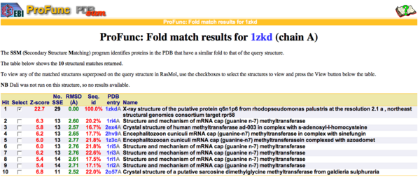
By using ProFunc (Laskowski et al, 2005) the most likely biochemical function of the unknown bacterial Protein 1zkd was determined as Methyltransferase. Matching structures were determined by SSM Secondary Structure Matching (Krissinel & Henrick, 2004) showing possible matches with 9 Methyltransferases from both human and bacteria (Fig.1).
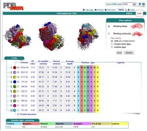
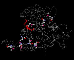
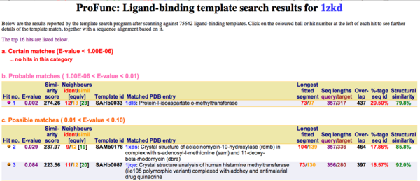
Ligand Template Matches LIG (Laskowski et al, 2005) revealed a probable match with the Protein-l-isoaspartate o-methyltransferase 1dl5 (Fig.2).
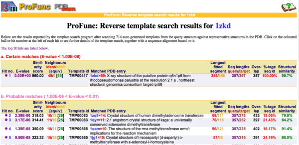
REV Reverse Template Matches (Laskowski et al, 2005) also showed probable matches for several methyltransferases (Fig.3).
Superfamily program searches against a library of Hidden Markov Models HMMs (Gough et al, 2001; Madera et al, 2004) derived from SCOP families revealed similarities to the superfamily S-Adenosylmethionine-dependent Methyltransferases (E-value 6.69e-06). No DNA binding motifs (helix-turn-helix) were found in the ProFunc search.
Genomic context of the 1izkd gene
Genomic context of 1zkd in the genome of Rhodopseudomonas palustris from the NCBI Entrez Gene database shows a genomic co-localisation with another transferase, an oxidase, a kinase and another hypothetical protein (Fig.4).

Localisation of 1zkd orthologs in the cell
Nucleo (Nuclear Protein Localisation Prediction) predicted a chance of 0.07 for the mouse ortholog and a chance of 0.20 for the human ortholog of 1zkd to be located in the nucleus (Hawkins et al, 2006).
LOCATE data was available for the mouse ortholog showing that it is a soluble, non-secreted protein with higher scores for a localisation in mitochondria or the cytoplasm (Fink et al, 2006).
Expression profiles of mouse and human orthologs
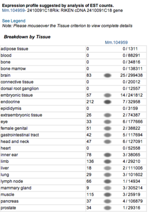
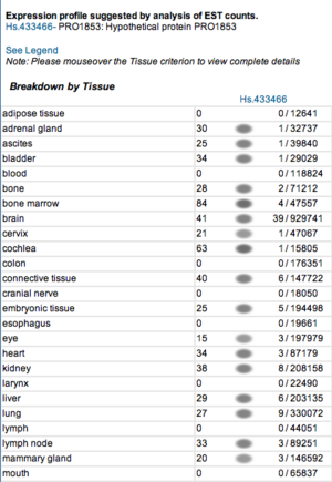
Expression profile data of the mouse and human ortholog were suggested by analysis of EST counts from NCBI UniGene database (http://www.ncbi.nlm.nih.gov/sites/entrez?db=unigene). ESTs were found in diverse tissues including brain, liver, lung, muscle and endocrine system showing that the target protein is expressed in a wide range of different cells (Fig.5a,b).
Electrostatic properties of the 1zkd protein surface
Electrostatic properties and surface charges of 1zkd were modelled using Adaptive Poisson-Boltzmann Solver APBS (Baker et al, 2001) and visualisation was performed by using Pymol (http://www.pymol.org). According to the resulting model, the 1zkd protein got a mostly negatively charged surface (Fig.6), indicating that interactions with the negatively charged backbone of nucleic acids are rather unlikely.
Evolution of Hypothetical Protein LOC55471 Isoform 1
BLAST P
BLASTP results produced 47 multiple aligned sequences to the bacterial sequence of the 1ZKD protein. The results are as follow BLASTP results.
The highlighted sequences show the 1ZKD protein sequence data and the closest match or best aligned sequence (>gi|39937419|ref|NP_949695.1| DUF185 [Rhodopseudomonas palustris CGA009]).
Clustal X
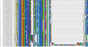
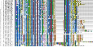
The following are some ClustalX images that were produced.
Protdist
Protdist results and how it looks like.
The number at the top of the page indicates the number of sequences uploaded.
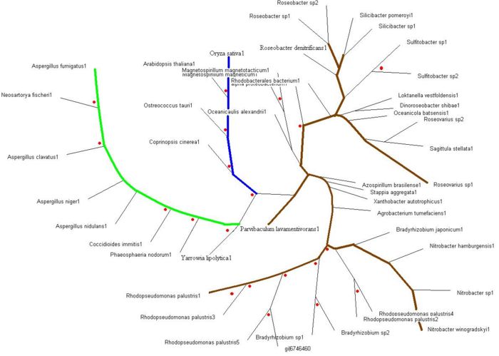
Evolutionary Tree
Return to Report on 1zkd
