ATP binding domain 4 Discussions
Discussion on Structure
IRU8 is a protein with an unknown function. The crystallization of 1RU8 was done from Pyrococcus furiosus that is expressed in Escherichia Coli with resolution of 2.7 Angstroms and an R-value of 0.218. Artificial chemical ligand, TRS(2-AMINO-2-HYDROXYMETHYL-PROPANE-1,3-DIOL) was used in the crystallization in order to maintain the integrity of the structure when crystallized. There is no actual ligand of this protein based on the the literature paper and from the Protein Data Base (PDB). This is probably because the function of the protein is still unknown. Determination of the ligand can also leads to the possible function of the protein. Hence, deduction of the ligand of this protein can only be done, based on other protein's ligand that has similar function or structure.
From the structure obtained from PyMOL, visualization of the secondary structure provide information on the surface properties covering positively charged regions and negatively charged regions (figure 3), its domains (figure 2.0) as well as ligand binding sites and surface clefts (figure 7.0), and also conservation of residues across different species(figure 2.0). It was observed that detailed secondary structure can be determined using PDBsum where conserved residues, protein motif and possible ligand binding site was highlighted (Figure 2). Information from the PDB and SCOP has indicated that 1RU8 is from the family of N-type ATP pyrophosphatases and also under the clan of PP-loop which is the strongly conserved motif Ser(12),Gly(13),Gly(14),Lys(15) and Asp(16) at the N terminus (Bork & Koonin 1994). This PP-loop was significant as it is the most conserved and is involved in ligand bindings and thus is likely to give function to our protein. Besides that, From CATH domain database, two main domains were found in 1RU8. The first domain ranges from residue 3-97 while the second domain residues from 98-232. Topology of Domain 1 is known to be of Rossmann A-B-A fold, and the superfamily of PP-loop is presence in this domain. Domain 2 is A-B complex classified in CATH.
Structural alignment via DALI enabled us to obtained proteins that is similar in terms of structure relatedness to 1RU8. Based on the DALI output, 2d13 is rejected since it is a hypothetical protein ph1257 that has no known function to compare to. Thus, 2NZ2 and 3BL5 was chosen since it is the most similar based on the z-score (Figure4). Information on 2NZ2 and 3BL5 by InterPro indicated that both protein has conserved motif of PP-loop at the N terminus which is similar to our protein. Hence, emphasizing again the importance of PP-loop to 1RU8's function. These 2 protein were analyzed based on the structural alignment and cleft size and volume. We inferred that the protein that has the most similar features to 1RU8 will probably gives similar function to 1RU8.
Structural alignment of 1RU8 with 2NZ2 and 3BL5 (Figure 5 and 6) show that 1RU8 and 2NZ2 has the most similar alignment compared to 1RU8 and 3BL5. Since 2NZ2 has a known function which is to catalyzes the citrulline and aspartate into argininosuccinate and pyrophosphate via hydrolysis of ATP, we inferred that the function of our protein may have similar mechanisms to 2NZ2 particularly the hydrolysis of ATP mechanisms. Based on Figure 5, we can observed that the ATP is located near the PP-loop highlighted in blue and Citrulline(yellow) is quite far from the loop which is supporting the fact that ATP is hydolysed for citrulline to activate. Hence, similar mechanism may be imply to 1RU8. Besides that, the active site or the binding site structure of both protein is quite similar(based on the structural alignment), and thus we also inferred that the substrate for 1RU8 must then have similar properties and organization of 2nz2 substrate (ATP and Citrulline) in a certain extent. However, the alignment is not convincingly similar due to domain B of 1RU8 (figure 5) that probably is the distinction of differences in function to 2NZ2.
Pockets and cavities in the structure is often associated with binding sites and active sites of proteins. Moreover, it is also believed that there is high possibility that the largest cavity is the active site with some exceptions (Liang et al. 1998). Shape and size parameters of protein pockets and cavities are thus are important for active site analysis. Identification and measurements of surface accessible pockets as well as interior inaccessible cavities of 1RU8, 2NZ2 and 3BL5 were obtained from CASTp (Figure 7.0 to 7.2).IT is inferred that the cleft volumes in proteins are related to their molecular interactions and functions (Laskowski et al. 1996). It was observed from the result, cleft of 1RU8 is quite similar in size and volume to 2NZ2 which suggesting that 1RU8 ligand may have similar size to 2NZ2 ligand. Thus, supporting the hypothesis that 1RU8 substrate or ligand may have similar properties to 2NZ2 substrate.
Despite the comparison of the two proteins to 1RU8, further structural comparison is needed in order for us to deduces other possible functions. Besides that determination of actual ligand based on experimental data may also help in finding the function of 1RU8.
Discussion on Function
From our findings, since both argininosuccinate synthetase (AS) and ATP binding domain 4 has striking similarities in terms of highly conserved PP-loop motif and cleft volume, it is possible to infer the mechanism of our protein based on the well-known AS reaction mechanism.
From the figure 6.1, Step 1. Argininosuccinate synthetase releases inorganic pyrophosphate after the formation of activate citrulline-adenylate. Step 2. Aspartate (the lone pair of N from amino group) undergoes nucleophilic attack on the carbonyl group on the activated citrulline-adenylate, hence forming argininosuccinate together with the release of AMP.
Therefore,
(1) Structure modelling with argininosuccinate synthetase (AS).
Our protein should be also involved in catalysing a substrate adenylation (means forming a phosphodiester bond between an amino acid and the phosphate group of AMP (adenosine monophosphate nucleotide) / (process of forming an adenylate, the salt or an ester of AMP) so as to activate a carbonyl or carboxyl group. (Lemke & Howell 2001) This activation is to facilitate the subsequent attack of a nitrogen-containing nucleophile from the substrate.
The structure of AS is used for the modelling ATP binding into our structure. The b and g phosphate groups of ATP are oriented by characteristic residues of the PP-loop, and then forming a salt linkage between the g-phosphate and the N atom of lysine. (Lemke & Howell 2001)
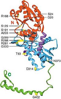
In the AS model in E. coli., R168 (see figure 6.2) has been proposed for pyrophosphate binding. The carbonyl oxygen of the second residue preceding the PP motif in AS (Ala-16), falls within the hydrogen bonding distance of the O2’ hydroxyl oxygen of the ribose. (Lemke & Howell 2001). Therefore, the alanine residue closely after the PP-motif in our protein, Ala(20) should be involved in stabilising ribose by forming H-bond with its O2’ hydroxyl oxygen. This may explain why relatively majority of aliphatic residues are conserved in the biggest gap in our cleft analysis (see //Bindsites2.PNG//) and relatively highly conserved detected by PDBsum.
It was proposed that the amide oxygen of a glutamine (Q46) should be involved in interacting with the N6 nitrogen of adenine. Relatively highly conserved Q104 in our structure (detected by PDBsum) might also be involved in similar interaction.
The most intriguing part is in the highly conserved PP-loop motif ([A/S]-[F/Y]-S-G-G-[L/V]-D-T-[S/T]) contains two absolutely conserved glycine residues, Gly(13) and Gly(14). In AS model, the two conserved glycine residues showed significantly different conformations in its uncomplexed and complexed structures, which are suspected to play a role in binding and release of pyrophosphate (PPi). (Lemke & Howell 2001) This agrees with glycine, lacking side chains, can provide a high degree of flexibility. Therefore, Gly(13)Gly(14) in ATP binding domain 4 should provide a large anion hole required for pyrophosphate binding. Lemke and Howell (2001) proposed that other N-type ATP pyrophosphatases models, if having glycine residues replaced, the steric hindrance created will crash with the bridge oxygen of the bound pyrophosphate.
The final residue of the PP motif in our structure, the hydroxyl group of Ser(17) should be involved in forming hydrogen bonds with the highly conserved residues, Gly(14) and Arg(60), meaning Ser(17) residue is important both for pyrophosphate binding and structural support by interacting with a helices, therefore H1 and H2 can be linked together. (see Figure 2.0 showing H1 and H2 in PDBsum)
// Ask Dan to highlight important residues, is it reasonably positioned to agree with the proposed catalytic mechanism? For Labelling help see p.10 of Lemke & Howell//
(2) Protein conformational change existed in the catalytic cycle.
The relative positions of all 3 substrates in AS suggest a strong requirement for a conformational change during catalysis. Our analysis of AS with substrates interactions indicates that PP-loop closely binds with ATP and is distantly related to citrulline (the substrate). This implies that as for our protein, only PP-loop is directly interacting with ATP, and its substrate may interact with ATP in the opposite end.
(3) Only domain A was involved in ATP-substrate binding; domain B suggested functions otherwise.
Domain B in our protein is poorly aligned with AS but closely resembled only in PP-loop-containing domain A region, which has been previously confirmed in our PyMOL alignment. It suggested that domain B of ATP binding domain 4 may be involved in substrates recognition which explains the subtle differences between our protein with AS. This hypothesis was based on our findings that the starting residues of domain B formed a hairpin-like structure that pointed to the core of ATP binding domain 4 that might suggest an intermolecular cooperation.
Future research should be focused on developing the crystallography of ATP binding domain 4 binding with its substrates. Attempts of building a cocrystallized structure with ATP and enzyme is often hard. Taking Lemke and Howell (2001)’s study as an example, cocrystallizing ATP with AS from E. coli. is detrimental which results into a poorly diffracting, easily cracked and dissolving crystals. Therefore, our study focuses only on inferring structure based on other N-type ATP pyrophosphatases. However, building a crystallised model will definitely provide us with insight to the enzymatic mechanism.
More structural and functional deduction experiments should be done based on site-directed mutagenesis on PP-loop and potential substrate-interacting residues in the groove identified in our structural analysis. More studies should be done based on elucidating domain B of ATP binding domain 4 which may be important for substrate selectivity.
Further research can be done on comparing the ATP binding domain 4 between human and Pyrococcus furiosus and look for any mutations of the structurally and catalytically important residues suggested in this study are associated with any ATP-related genetic diseases.
Lemke, C, T. and Howell, P.L. (2001), The 1.6A Crystal Structure of E.coli Argininosuccinate Synthetase Suggests a Confomational Change during Catalysis, Structure 9:1153-1164.

