ATP binding domain 4 Structures: Difference between revisions
From MDWiki
Jump to navigationJump to search
Anharmustafa (talk | contribs) No edit summary |
Anharmustafa (talk | contribs) No edit summary |
||
| Line 20: | Line 20: | ||
Secondary Structure and Location of P-loop (ATP binding site) | Secondary Structure and Location of P-loop (ATP binding site) | ||
1RU8 has 2 domain | |||
[[Image:1RU8 as 2 domain.png]] | |||
Revision as of 04:58, 23 May 2009
Daniel's page in progress
Useful info
Here is some useful paper. check it out Link to Paper: http://pfam.sanger.ac.uk/family?acc=PF00764 Dali: http://ekhidna.biocenter.helsinki.fi/dali_server/results/20090512-0051-3897fbdf5431ad2e808e66dde1070610/index.html#alignment-7 Interpro: http://www.ebi.ac.uk/interpro/DisplayIproEntry?ac=IPR002761
SCOP Work
http://scop.mrc-lmb.cam.ac.uk/scop/data/scop.b.d.cj.c.b.bb.html
Pymol Work
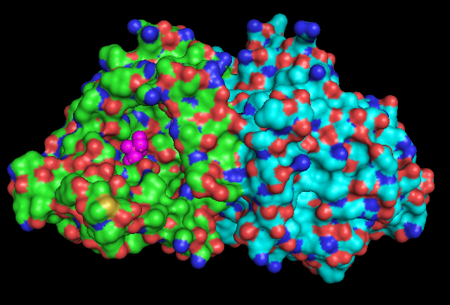
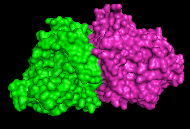
Surface Structure
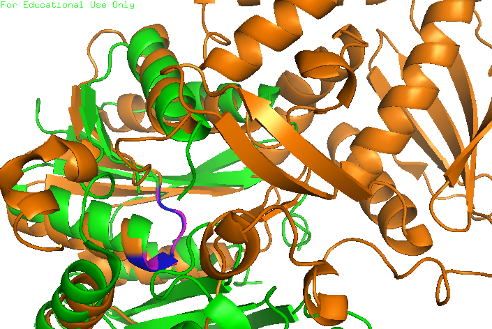
Secondary Structure and Location of P-loop (ATP binding site)
1RU8 has 2 domain
align (align 1RU8 & i. 11-16, 2NZ2 & i. 11-15)


