ATP binding domain 4 Structures: Difference between revisions
Anharmustafa (talk | contribs) No edit summary |
Anharmustafa (talk | contribs) No edit summary |
||
| Line 33: | Line 33: | ||
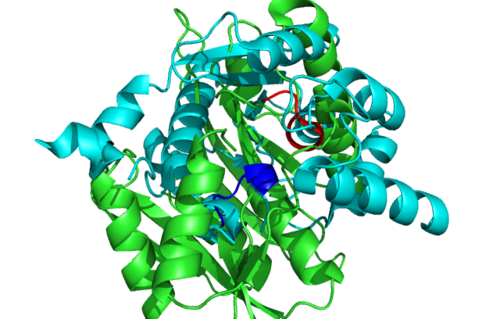
== 1RU8 is a dimer == | == 1RU8 is a dimer == | ||
[[Image:1RU8 as 2 domain.png | [[Image:1RU8 as 2 domain.png]] | ||
== Secondary Structure and Location of P-loop (ATP binding site) == | == Secondary Structure and Location of P-loop (ATP binding site) == | ||
Revision as of 14:35, 1 June 2009
General information
General information from PDB indicates that :
(a) 1RU8 is a putative n-type ATP pyrophosphatase isolated from Pyrococcus furiosus, expressed in Escherichia Coli.
(b) Is a member of clan PP-loop
(c) Resolution of 2.7 angstroms, with an r-value of 0.218.
(d) Ligand chemical component identified as TRS (2-AMINO-2-HYDROXYMETHYL-PROPANE-1,3-DIOL).
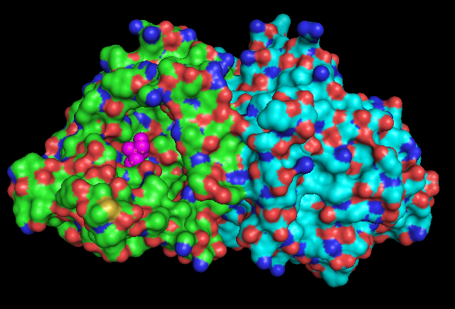
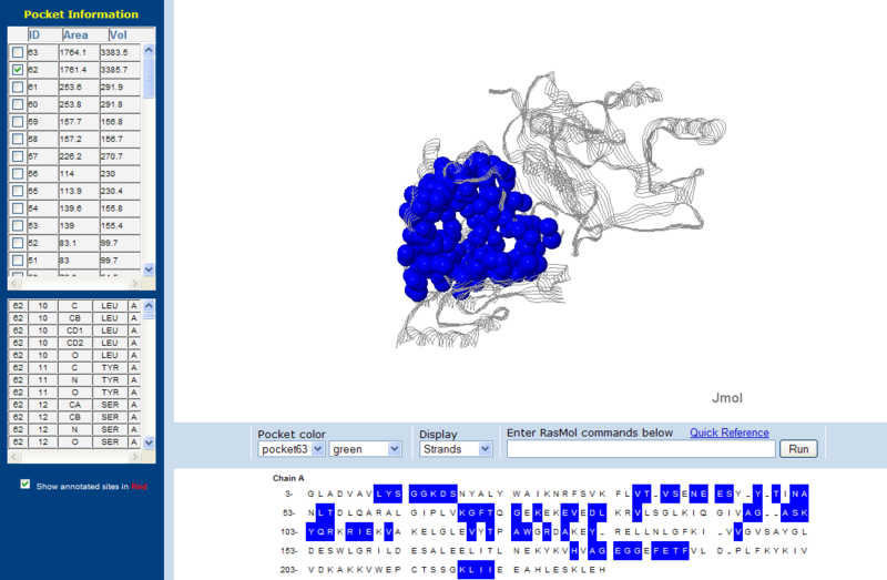
Surface Structure
Pymol Visualization
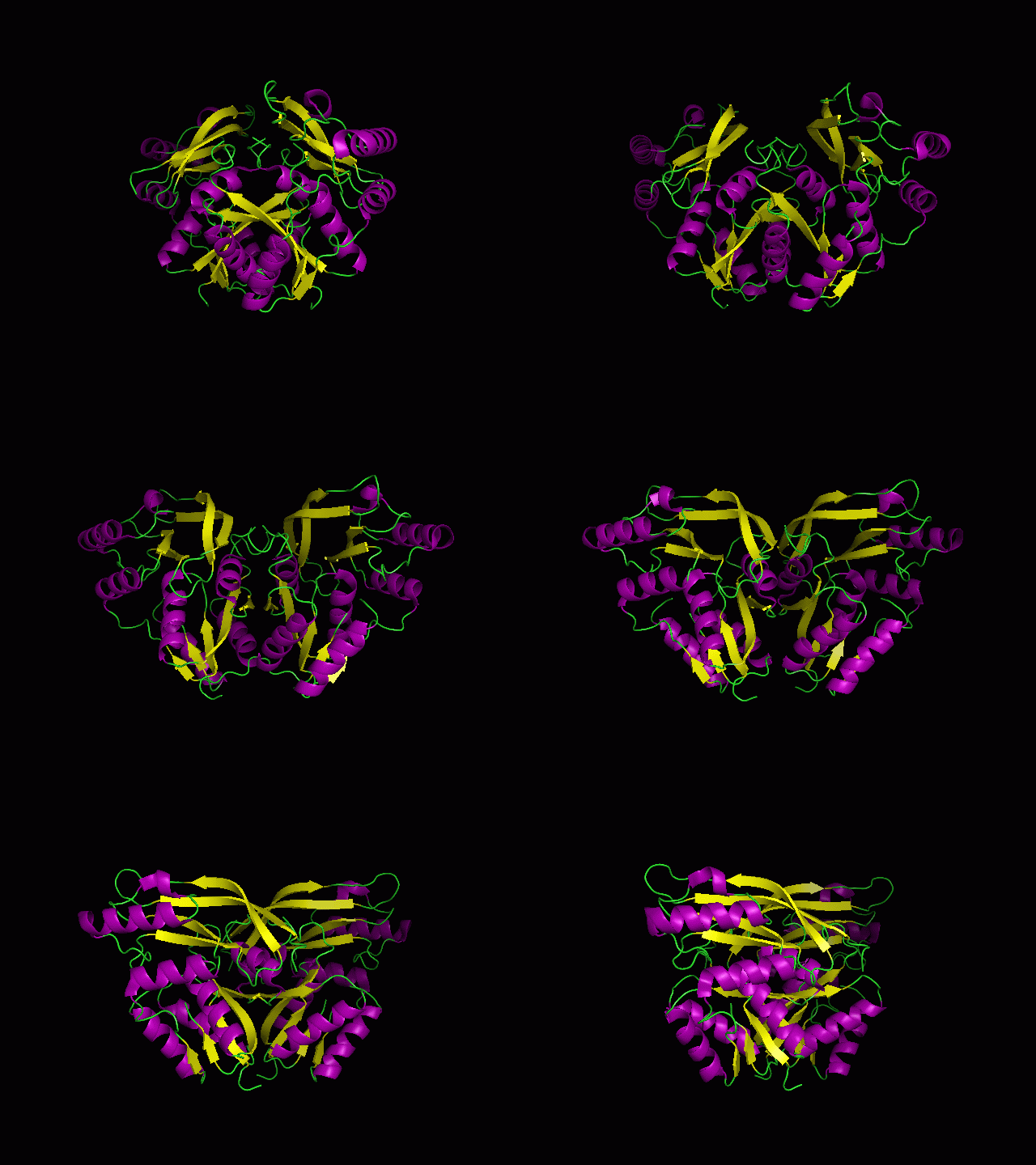
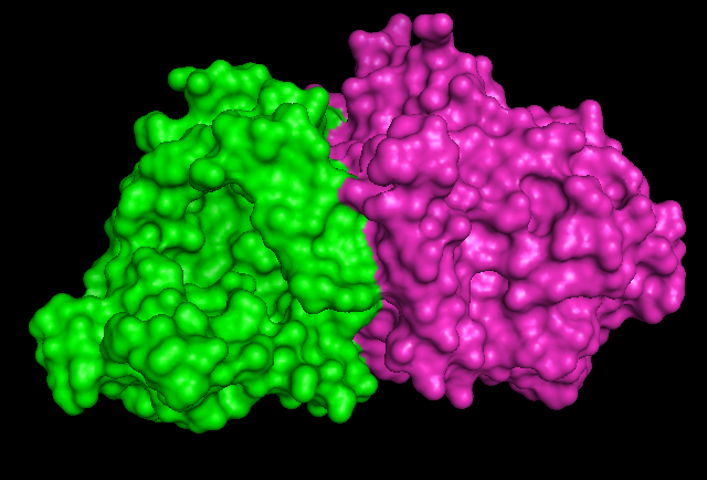
1RU8 is a dimer
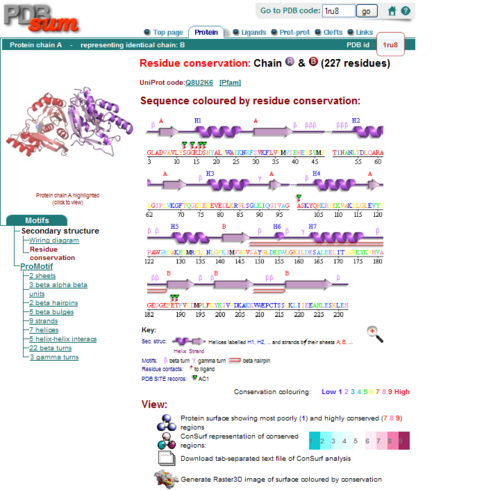
Secondary Structure and Location of P-loop (ATP binding site)
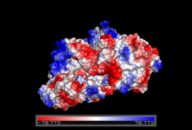
Electrostatic Surface Potential
Structure Similarities
Z score , the statistical significance of the similarity between protein-of-interest and other neighbourhood proteins. The program optimizes a weighted sum of similarities of intramolecular distances.
Root Mean Square Distance (RMSD), root-mean-square deviation of C-alpha atoms in the least-squares superimposition of the structurally equivalent C-alpha atoms. RMSD is not optimized and is only reported for information.
lali, the number of structurally equivalent residues.
nres, or the total number of amino acids in the hit protein.
%id, percentage of identical amino acids over structurally equivalent residues.
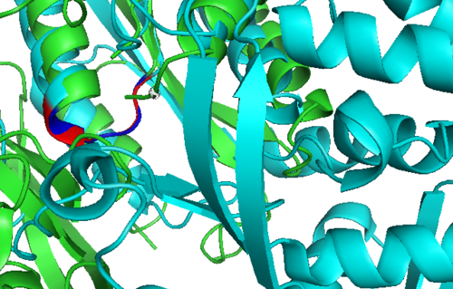
Structure Alignment
1RU8 and 2NZ2
1RU8 and 3BL5
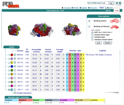
Surface Clefts
Surface Topography
