Coenzyme A Synthase's Relation To Other Structurally Related Proteins: Difference between revisions
From MDWiki
Jump to navigationJump to search
No edit summary |
No edit summary |
||
| Line 4: | Line 4: | ||
|- | |- | ||
| | | | ||
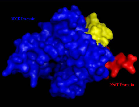
[[Image:Domains_2F6r.jpg|thumb| | [[Image:Domains_2F6r.jpg|thumb|450px|Surface structure of ''Mus musculus'' Coenzyme A Synthase with Phosphopantetheine adenylyltransferase (PPAT) and Dephospho-CoA kinase (DPCK) domains marked in red and blue respectfully.|none]] | ||
|- | |- | ||
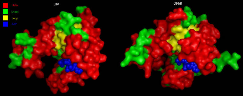
|colspan="2"|[[Image:COASYSecondary structure comparsion.jpg|thumb|800px|'''Figure 5 '''<BR> Structural comparison of Dephospho-COA Kinase (IJJV) and Bifunctional coenzyme A synthase from ''Mus musculus'' (2F6R) with bound ATP.|none]] | |colspan="2"|[[Image:COASYSecondary structure comparsion.jpg|thumb|800px|'''Figure 5 '''<BR> Structural comparison of Dephospho-COA Kinase (IJJV) and Bifunctional coenzyme A synthase from ''Mus musculus'' (2F6R) with bound ATP.|none]] | ||

