DHRS1 Results
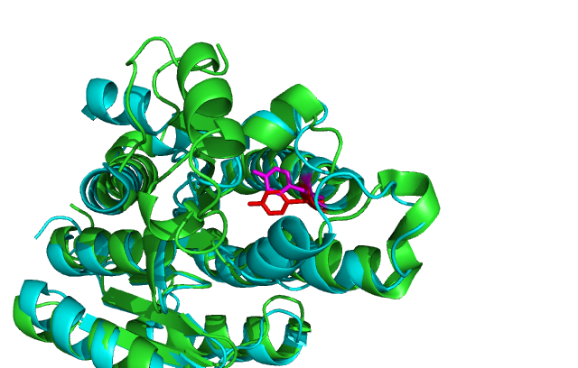
Results/structure Dehydrogenase/reductase SDR family member 1 has highly conserved structure compared across the SDR family.
SDR family is part of the Super Family; NAD(P)-binding Rossmann-fold domain proteins all of which have the Rossmann-fold domain, which is characterised a central β-sheet surrounded by α-helicies. This buts them in the Alpha and Beta proteins (α/β) class.
Some key residues are conserved across the entire family. Notable a Tyrosine that binds NADPH. It is part of a larger motif (SxxxxxxxxxxxxYxxxK) that includes two other residues involved in binding NADPH.
There is another highly conserved motif (LDVLD) involved in the initial folding of the protein and forms part of the hydrophobic core.
BLAST
The protein sequence for the SDR family member 1 protein being investigated was downloaded from the PDB site, using the accession code 2qq5. This was then iteratively searched in blastp using an offline, non redundant copy of the database produced on the 28th of April 2008.
MULTIPLE SEQUENCE ALIGNMENT
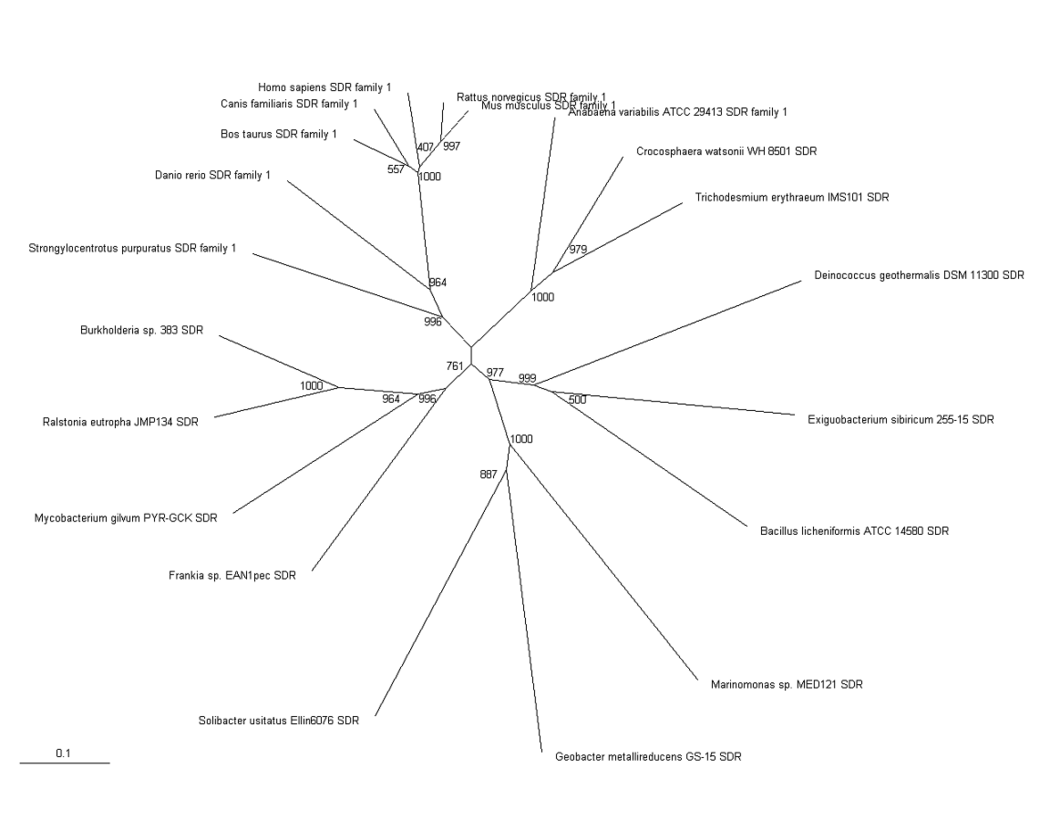
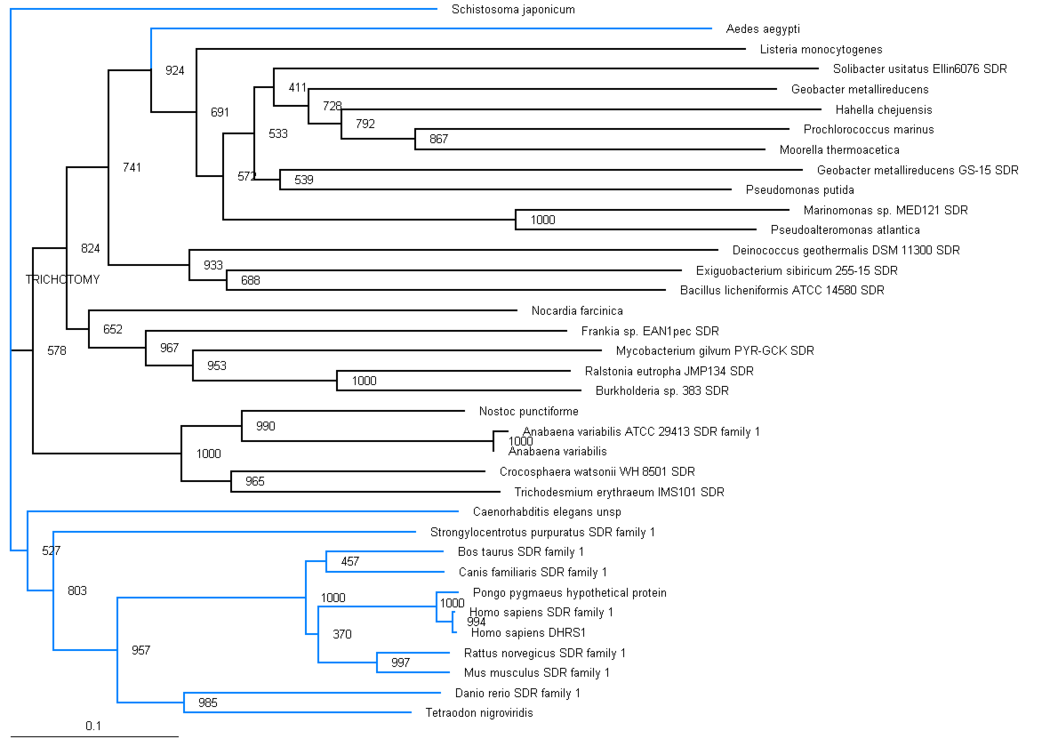
The results from the blast search were then screened and a selection was of these results were used for a multiple sequence alignment using ClustalX. This result was boostrapped and these values checked and more sequences were added to improve the resolution of specific branches. A bootstrapped phylogram was produces, aswell as a radial tree.
Table 1
PDB/chain identifiers and structural alignment statistics for DALI search
No:	Chain	Z	rmsd	lali	nres	%id	Description
1:	2qq5-A	48.1	0.0	238	238	100	MOL:DEHYDROGENASE/REDUCTASE SDR1;
2:	2uvd-A	29.6	2.1	220	246	27	MOL: 3-OXOACYL-(ACYL-CARRIER-PROTEIN)  REDUCTASE;
3:	1yde-F	29.3	2.1	216	256	29	MOL: RETINAL DEHYDROGENASE/REDUCTASE 3;
4:	1vl8-B	29.1	2.0	220	252	27	MOL: GLUCONATE 5-DEHYDROGENASE;
5:	2bgk-A	29.0	2.1	219	267	25	MOL: RHIZOME SECOISOLARICIRESINOL DEHYDROGENASE;
6:	2q2q-D	28.9	2.0	217	255	26	MOL: BETA-D-HYDROXYBUTYRATEDEHYDROGENASE;
7:	1rwb-F	28.8	2.2	221	261	24	MOL: GLUCOSE 1-DEHYDROGENASE;
8:	1rwb-A	28.8	2.3	222	261	24	MOL: GLUCOSE 1-DEHYDROGENASE;
9:	1gee-A	28.8	2.3	222	261	24	MOL: GLUCOSE 1-DEHYDROGENASE;
10:	1gco-A	28.8	2.3	222	261	24	MOL: GLUCOSE DEHYDROGENASE;
11:	2zat-A	28.7	2.1	221	251	23	MOL: DEHYDROGENASE/REDUCTASE SDR4;
12:	1gee-B	28.7	2.3	221	261	24	MOL: GLUCOSE 1-DEHYDROGENASE;
13:	1gco-E	28.7	2.3	221	261	24	MOL: GLUCOSE DEHYDROGENASE;


