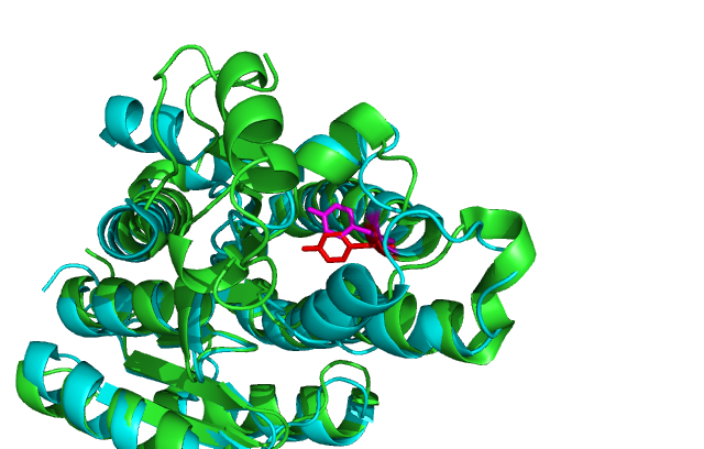
DHRS1 Results
Results/Evolution
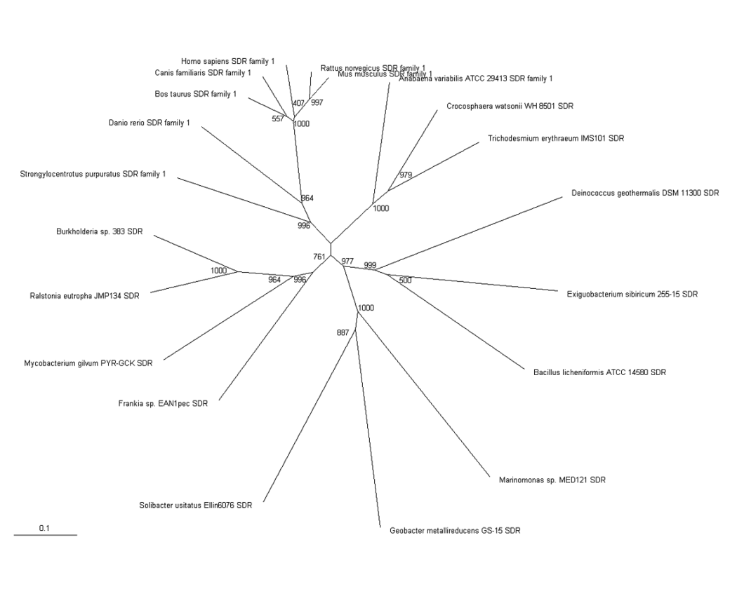
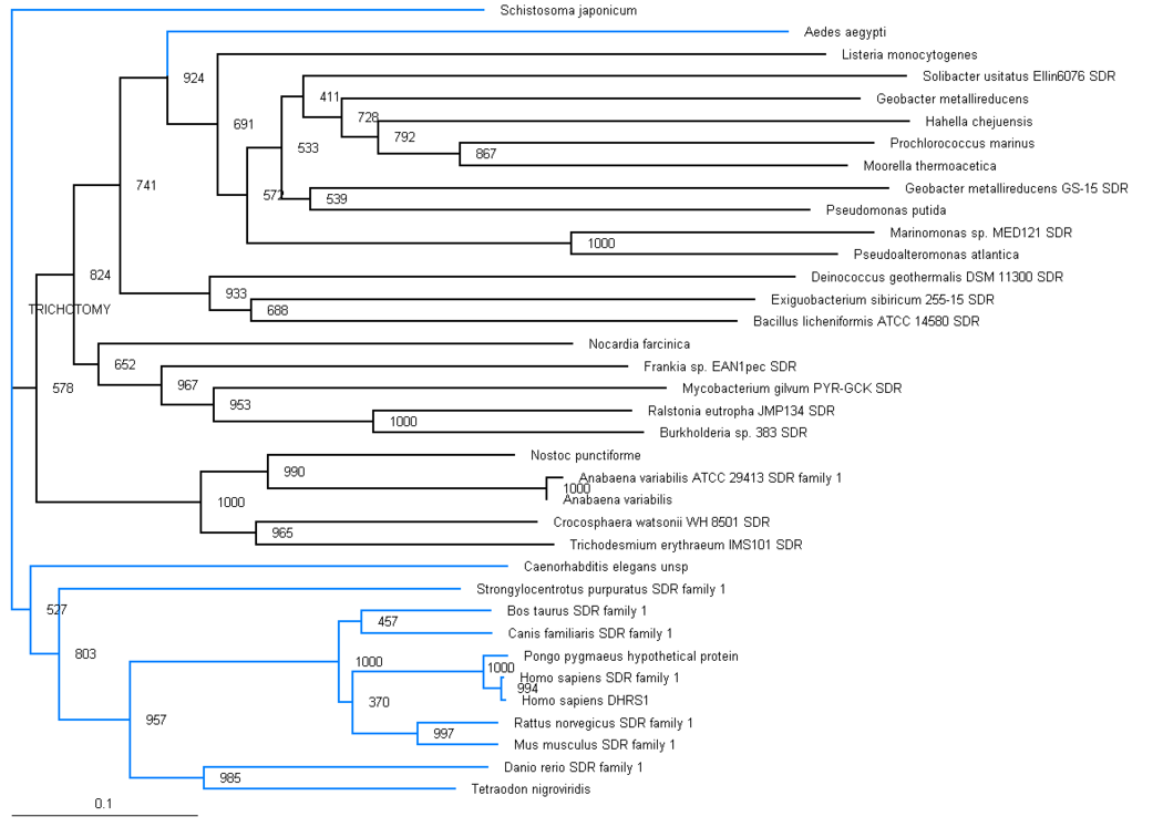
The phylogenic tree shows a small section of the family is contained in land based higher organisms (Bos taurus through to Mus Muluscus), a smaller section is found in sea based eukaryotes (Danio rerio and Tetraodon nigroviridis) and the majority of the family is round in bacteria. The majority of bacteria found to possess this family are proteobacteria, although there are also gram positive rod (eg. bacillus) and cyanobacteria (eg. anabena). This suggests that this particular family has been present for a long time and has evolved from bacteria, most likely proteobacteria.
Results/structure
DHRS1 contain 10 α helices 1 central β-sheet region.
Structure comparison
Comparison of DHRS1 to 3-OXOACYL-(ACYL-CARRIER-PROTEIN) REDUCTASE its closest structurally related protein, finds that whilst it shares only 27% of its sequence, it is structurally very similar (Fig 2). They share many structural features including the central β-sheet region and many α helices. Key conserved residues such as the tyrosine shown are also in very similar positions eluding to a similar function.
Table 1
PDB/chain identifiers and structural alignment statistics from DALI search
No:	Chain	Z	rmsd	lali	nres	%id	Description
1:	2qq5-A	48.1	0.0	238	238	100	MOL: DEHYDROGENASE/REDUCTASE SDR1;
2:	2uvd-A	29.6	2.1	220	246	27	MOL: 3-OXOACYL-(ACYL-CARRIER-PROTEIN)  REDUCTASE;
3:	1yde-F	29.3	2.1	216	256	29	MOL: RETINAL DEHYDROGENASE/REDUCTASE 3;
4:	1vl8-B	29.1	2.0	220	252	27	MOL: GLUCONATE 5-DEHYDROGENASE;
5:	2bgk-A	29.0	2.1	219	267	25	MOL: RHIZOME SECOISOLARICIRESINOL DEHYDROGENASE;
6:	2q2q-D	28.9	2.0	217	255	26	MOL: BETA-D-HYDROXYBUTYRATEDEHYDROGENASE;
7:	1rwb-F	28.8	2.2	221	261	24	MOL: GLUCOSE 1-DEHYDROGENASE;
8:	1rwb-A	28.8	2.3	222	261	24	MOL: GLUCOSE 1-DEHYDROGENASE;
9:	1gee-A	28.8	2.3	222	261	24	MOL: GLUCOSE 1-DEHYDROGENASE;
10:	1gco-A	28.8	2.3	222	261	24	MOL: GLUCOSE DEHYDROGENASE;
11:	2zat-A	28.7	2.1	221	251	23	MOL: DEHYDROGENASE/REDUCTASE SDR4;
12:	1gee-B	28.7	2.3	221	261	24	MOL: GLUCOSE 1-DEHYDROGENASE;
13:	1gco-E	28.7	2.3	221	261	24	MOL: GLUCOSE DEHYDROGENASE;
