File:PsaA docked to E-cadherin in simulation cell.png
From MDWiki
Jump to navigationJump to search
Size of this preview: 665 × 599 pixels. Other resolution: 1,044 × 941 pixels.
Original file (1,044 × 941 pixels, file size: 739 KB, MIME type: image/png)
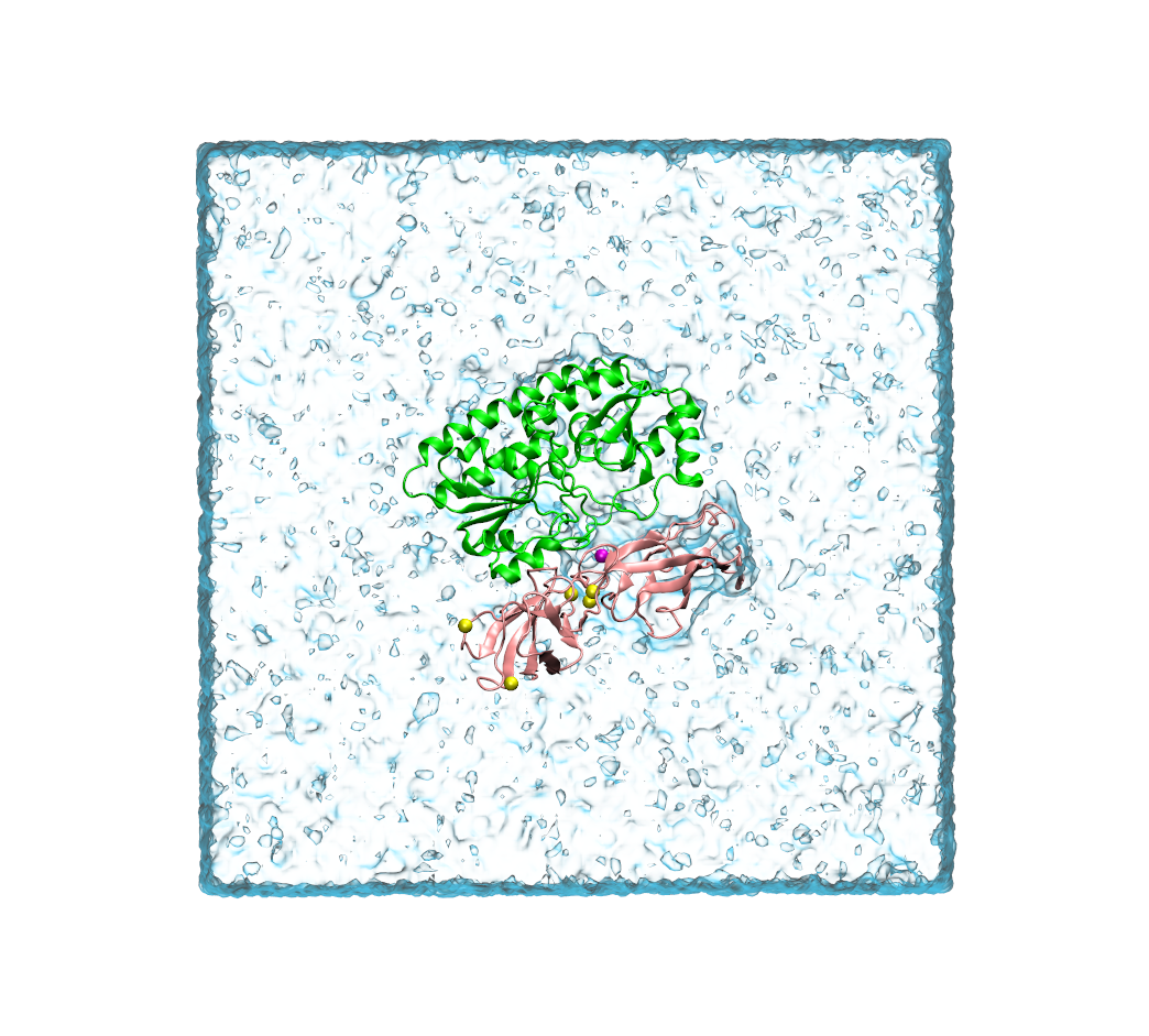
Pneumococcal surface adhesin A (PsaA) is docked to one of the two extracellular domains of an E-cadherin monomer. It is suspended in a box of water. The image is a snapshot that was taken using VMD. It shows the two proteins in cartoon representation, and calcium ions and a model metal ion as yellow and magenta spheres, respectively.
File history
Click on a date/time to view the file as it appeared at that time.
| Date/Time | Thumbnail | Dimensions | User | Comment | |
|---|---|---|---|---|---|
| current | 04:06, 13 March 2015 | | 1,044 × 941 (739 KB) | Uqbcaron (talk | contribs) | Pneumococcal surface adhesin A (PsaA) is docked to one of the two extracellular domains of an E-cadherin monomer. It is suspended in a box of water. The image is a snapshot that was taken using VMD. It shows the two proteins in cartoon representation, ... |
You cannot overwrite this file.
File usage
There are no pages that use this file.