|
|
| Line 109: |
Line 109: |
| **N-acetylneuraminic acid phosphatase orthologs shared 3 motifs found in phosphatases | | **N-acetylneuraminic acid phosphatase orthologs shared 3 motifs found in phosphatases |
|
| |
|
| *HAD family | | |
| ** Phosphatase activity CO–P bond hydrolysis | | *HAD (Haloacid dehalogenase-like) family |
| ** Dehalogenase activity C–halogen bond hydrolysis | | ** Phosphatase activity: CO–P bond hydrolysis |
| ** Phosphonatase C–P bond hydrolysis | | ** Dehalogenase activity: C–halogen bond hydrolysis |
| ** Phosphoglucomutase CO–P bond hydrolysis and intramolecular phosphoryl transfer | | ** Phosphonatase: C–P bond hydrolysis |
| | ** Phosphoglucomutase: CO–P bond hydrolysis and intramolecular phosphoryl transfer |
|
| |
|
|
| |
|
| phosphatase, phosphoglucomutase, phosphonatase, and dehalogenase activities 6. HAD-like hydrolases represent the largest family of predicted small molecule phosphatases encoded in the genomes of bacteria, archaea, and eukaryotes, with 6,805 proteins in data bases 7. HADs share little overall sequence similarity (15–30% identity), but they can be identified by the presence of three short conserved sequence motifs 7. Most of the characterized HADs have phosphatase activity (CO–P bond hydrolysis), catalyze dehalogenase activity (C–halogen bond hydrolysis), phosphonatase (C–P bond hydrolysis), and phosphoglucomutase (CO–P bond hydrolysis and intramolecular phosphoryl transfer) reactions 6.
| |
|
| |
|
|
| |
|
| Line 125: |
Line 125: |
| ** Gene map locus 20p11 | | ** Gene map locus 20p11 |
|
| |
|
| * Main form of sialic acid in vertebrates
| |
| ** Important roles in protein-protein and cell-cell recognition
| |
|
| |
|
| [[Image:Document2_01.png]] | | [[Image:Document2_01.png]] |
| Line 133: |
Line 131: |
|
| |
|
|
| |
|
| ---- | | * Main form of sialic acid in vertebrates |
| | | ** Important roles in protein-protein and cell-cell recognition |
| , illustrate that 2gfh is a hydrolase. Profunc searches (Figure 17) on 2gfh also
| | ** Dependent on the presence of Mg<sup>2+</sup> |
| | | ** Inhibited by vanadate and Ca<sup>2+</sup> |
| show that it possesses hydrolase activity. The highest score for Gene Ontology (Figure 17) states it used for metabolism and possesses
| |
|
| |
|
| phosphoglycolate phosphatase activity. Hydrolyase is an enzyme which catalyzes hydrolysis reaction (Figure 18), which is the addition of the
| |
|
| |
|
| hydrogen and hydroxyl ions of water to a molecule with its consequent splitting into two or more simpler molecules. Hydrolase is the systematic
| |
|
| |
|
| name for any enzyme of EC class 3.
| |
|
| |
|
| | <font size = "4">'''Role in Bacteria - ''E.coli'''''</font> |
|
| |
|
| | * Hydrolyze a wide range of phosphorylated metabolites |
| | ** Carbohydrates |
| | ** Nucleotides, |
| | ** Organic acids |
|
| |
|
| '''Figure 18. '''Hydrolyase catalyze the hydrosis of the chemical bond between A and B, resulting of 2 simple molecules.
| | * Kuznetsova ''et al'' - glycolysis and pentose phosphate pathway |
| | | ** Fructose-1-phosphate |
| | | ** Glucose-6-phosphate |
| ----
| | ** Mannose-6-phosphate |
| | | ** 2-deoxyglucose-6-phosphate |
| | | ** Fructose-6- phosphate |
| [[Image:Document9_01.png]]
| | ** Ribose-5-phosphate |
| | | ** Erythrose- 4-phosphate |
| The MSA for the query sequence and the other 35 sequences shows several conserved motifs. The 1<sup>st</sup> conserved motif
| |
| | |
| consists of almost invariant region of aspartic acid (D), only the 33<sup>rd</sup> protein (gi: <nowiki>|</nowiki>45552117<nowiki>|</nowiki>)
| |
| | |
| showing gap. The 2<sup>nd</sup> motif shows conserved and invariant of leucine (L), threonine (T), asparagine (N) and glycine (G). The
| |
| | |
| 3<sup>rd</sup> motif shows 2 invariant amino acid residues of lysine (K), proline (P), valine (V), glycine (G), aspartic acid (D) and
| |
| | |
| isoleucine (I). This correlates with the study done by Maliekal ''et al'' and strongly suggested that the query protein is a phosphatase.
| |
| | |
| | |
| MSA of the query protein Neu5Ac phosphatase with 35 others proteins. Only the 60<sup>th</sup> – 70<sup>th</sup> and the
| |
| | |
| 210<sup>th</sup> -300<sup>th</sup> amino acid sequence were shown to illustrate the conserved and invariant regions. The 3 boxed-up sequences
| |
| | |
| were either conserved or invariant regions.
| |
| | |
| | |
| | |
| [[Image:Document17_01.png]]
| |
| [[Image:Document17_03.png]]
| |
| '''Figure 17. (A)''' List of all matched protein name terms for 2gfh.''' (B) '''List of all matched Gene Ontology terms for 2gfh. The score in
| |
| | |
| red is a measure of how strongly the term is predicted from the hits obtained by the different methods. The scores in blue show each
| |
| | |
| method<nowiki>’</nowiki>s contribution to the total score (with the number of relevant sequences/structures shown in brackets in grey).
| |
| | |
| (http://www.ebi.ac.uk/thornton-srv/databases/cgi-bin/pdbsum/)
| |
| | |
| | |
| | |
| | |
| ''E. coli ''HADs hydrolyze a wide range of phosphorylated metabolites, including carbohydrates, nucleotides, organic acids, and coenzymes.
| |
| | |
| Studies have shown that the most common substrates in metabolism such as glycolysis and pentose phosphate pathway (Figure 18). These enzymes
| |
| | |
| were fructose-1-phosphate, glucose-6-phosphate, mannose-6-phosphate, 2-deoxyglucose-6-phosphate, fructose-6- phosphate, ribose-5-phosphate, and
| |
| | |
| erythrose- 4-phosphate <sup>13</sup>.
| |
|
| |
|
|
| |
|
| Line 200: |
Line 160: |
| [[Image:Document20_02.png]] | | [[Image:Document20_02.png]] |
|
| |
|
| '''Figure 20. '''The schematic diagrams of glycolysis and pentose phosphate metabolic pathways. The green arrows show the substrates that are hydrolyzed by HADs '''(A)''' Glycolysis pathway with substrates that are hydrolyze by HADs: glucose 6-phosphate, fructose 6-phosphate and dihydroxyacetone phosphate. '''(B)''' Pentose phostphate pathway with substrates that are hydrolyze by HADs: glucose-6-phosphate, fructose-6-phosphate, dihydroxyacetone phosphate, glyceraldehyde-3-phosphate, gluconate 6-phosphate and erythrose-4-phosphate.
| | Schematic diagrams of glycolysis and pentose phosphate metabolic pathways. The green arrows show the substrates that are hydrolyzed by HADs |
| | |
| (http://www.steve.gb.com/science/core_metabolism.html)
| |
| | |
| | |
| =='''Proposed Functions'''==
| |
| | |
| * Hydrolase Activity
| |
| | |
| * Magnesium Ion Binding
| |
| | |
| * N-acylneuraminate-9-phosphatase Activity
| |
| | |
| * Phosphoglycolate Phosphatase Activity
| |
| | |
| | |
| | |
| | |
| | |
| [http://amigo.geneontology.org/cgi-bin/amigo/go.cgi?view=details&depth=1&query=0016787 1. Hydrolase Activity]
| |
| | |
| [http://amigo.geneontology.org/cgi-bin/amigo/go.cgi?view=details&depth=1&query=0000287 2. Magnesium Ion Binding]
| |
| | |
| [http://amigo.geneontology.org/cgi-bin/amigo/go.cgi?view=details&depth=1&query=0050124 3. N-acylneuraminate-9-phosphatase Activity]
| |
| | |
| [http://amigo.geneontology.org/cgi-bin/amigo/go.cgi?view=details&depth=1&query=0008967 4. Phosphoglycolate Phosphatase Activity]
| |
| | |
| | |
| | |
| {| border="1" cellpadding="15" cellspacing="0"
| |
| |+'''Table of functions'''
| |
| |Catalytic activity
| |
| |N-acylneuraminate 9-phosphate + H2O = N-acylneuraminate + phosphate
| |
| |-
| |
| |Cofactor
| |
| |Magnesium (By similarity)
| |
| |-
| |
| |Enzyme regulation
| |
| |Inhibited by vanadate and calcium (By similarity)
| |
| |-
| |
| |Pathway
| |
| |Carbohydrate metabolism; aminosugar metabolism
| |
| |-
| |
| |Similarity
| |
| |Belongs to the haloacid dehalogenase-like hydrolase superfamily. NANP family
| |
| |}
| |
| | |
| | |
| Using ProFunc: Ligand-binding template search results for 2gfh.
| |
| | |
| Structural similarity: 91.5%
| |
| | |
| E-value < 1.00E-06 ( 7.22E-07)
| |
| | |
| Similarity score: 364.02
| |
| | |
| PBD id: 2hi0
| |
| | |
| Name: Hydrolase
| |
| | |
| Title: Crystal structure of putative phosphoglycolate phosphatase (yp_619066.1) from lactobacillus delbrueckii subsp. Bulgaricus atcc baa-365 at 1.51 a resolution
| |
| | |
| Source: Lactobacillus delbrueckii. Bacteria. Gene: yp_619066.1. Expressed in: escherichia coli.
| |
| | |
| Reaction: 2-phosphoglycolate + H2O = glycolate + phosphate
| |
| | |
| [[Image:Tree.JPG|thumb|Description|left]]
| |
| | |
| | |
| | |
| | |
| | |
| | |
| | |
| | |
| | |
| | |
| | |
| | |
| | |
| | |
| | |
| | |
| | |
| | |
| | |
| | |
| =='''GO Terms'''==
| |
|
| |
| Polymer: haloacid dehalogenase-like hydrolase domain containing 4
| |
| | |
| Molecular Function: None
| |
| | |
| Biological Process: None
| |
| | |
| Cellular Component: None
| |
| | |
| | |
| =='''Function in Human'''==
| |
| | |
| haloacid dehalogenase-like hydrolase domain
| |
| | |
| (Found in OMIM)
| |
| | |
| Neu5Ac-9-PHOSPHATE PHOSPHATASE HALOACID DEHALOGENASE-LIKE HYDROLASE DOMAIN-CONTAINING PROTEIN-4; HDHD4
| |
| Gene map locus 20p11
| |
| | |
| | |
| Neu5Ac-9-phosphate phosphatase (Neu5Ac-9-Pase; EC 3.1.3.29) dephosphorylates Neu5Ac-9-phosphate to form N-acetylneuraminate (Neu5Ac), the main form of sialic acid in vertebrates that has important roles in protein-protein and cell-cell recognition.
| |
| | |
| | |
| CLONING
| |
| | |
| Maliekal et al. (2006) purified Neu5Ac-9-Pase from rat liver and isolated the phosphatase activity. Using SDS-PAGE analysis and tandem mass spectrometry, they identified a haloacid dehalogenase-like hydrolase domain containing-4 (HDHD4) protein. Purified recombinant human HDHD4 catalyzed the dephosphorylation of Neu5Ac-9-phosphate with a catalytic efficiency more than 2 orders of magnitude higher than for any other substrate tested. The 248-amino acid human HDHD4 protein has 3 motifs found in phosphatases of the haloacid dehalogenase (HAD) family: the first with 2 extremely conserved aspartates, the second comprising a conserved serine or threonine, and the third comprising a conserved lysine and 2 conserved aspartates.
| |
| | |
| | |
| GENE STRUCTURE
| |
| | |
| Maliekal et al. (2006) determined that the human HDHD4 gene contains 2 exons.
| |
| | |
| | |
| GENE FUNCTION
| |
| | |
| Maliekal et al. (2006) determined that the phosphatase activity of human Neu5Ac-9-Pase protein was dependent on the presence of Mg(2+) and was inhibited by vanadate and Ca(2+), which is characteristic of members of the HAD family of phosphatases.
| |
| | |
| | |
| | |
| | |
| | |
| | |
| | |
| ----
| |
| | |
| info
| |
| | |
| N-acetylneuraminic acid phosphatase
| |
| | |
| Homologous to mouse (Mus musculus)
| |
| | |
| Haloacid Dehalogenase Like Hydrolase Domain Containing 4
| |
| | |
| Classified as Hydrolase
| |
|
| |
|
| [http://www.ebi.ac.uk/thornton-srv/databases/profunc/index.html Infomation on ProFunc] (useful 2gfhA)
| |
|
| |
|
| [http://www.ebi.ac.uk/pdbsum/ Information on PDBsum] (2gfh)
| |
|
| |
|
| From [http://www.ebi.ac.uk/cgi-bin/sumtab?tool=fasta&jobid=fasta-20070515-07270763 FASTA]
| |
| most likely function is N-acetylneuraminic acid phosphatase.
| |
|
| |
|
| The haloalkanoate dehalogenase superfamily (HADSF) is one of the largest and most ubiquitous enzyme families identified to date, with over 3,000 members in organisms ranging from bacteria to humans. Remarkable diversity of chemistry and function has emerged through evolution of the HAD catalytic scaffold. Despite the name, the dehalogenases, which catalyze carbon group transfer, represent a minute fraction of the family. All other known catalytic activities are directed at phosphoryl transfer. Numerous proteins from the HADSF are found in each organism (29 in E. coli and 58 in humans, for example) where they perform a diverse collection of novel physiological functions in primary and secondary metabolism, membrane transport, signal transduction, and nucleic acid repair. http://biophysics.bumc.bu.edu/faculty/allen/allenpage/had.htm
| |
|
| |
|
| Haloacid dehalogenases (E.C.3.8.1.2) are members of the haloacid dehalogenase superfamily, which also contains ATPases, phosphatases and epoxide hydrolases. They catalyse the conversion of α-halo-carboxylic acids to the corresponding hydroxyalkanoic acid by nucleophilic attack on the α-carbon by a conserved aspartic acid residue to form an ester intermediate, which is then further hydrolysed by a water molecule. There are three subtypes of haloacid dehalogenase based on substrate specificity; those that can use both enantiomers as substrates, those that act only on the L enantiomer and those that act only on the D enantiomer.
| |
|
| |
|
| ---- | | ---- |
|
| |
|
| Press here to go [http://compbio.chemistry.uq.edu.au/mediawiki/index.php/BIOL3004_2007 ''Back''] | | Press here to go [http://compbio.chemistry.uq.edu.au/mediawiki/index.php/BIOL3004_2007 ''Back''] |
The predicted function based on the evolution and structure

Hydrolyase catalyze the hydrosis of the chemical bond between A and B, resulting of 2 simple molecules
- Hydrolase
- Catalyze hydrolysis reaction
- Addition of the hydrogen and hydroxyl ions of water
- Splitting into 2 or more simpler molecules
- EC class 3
Pfam
|
Hydrolase: domain 1 of 1, from 18 to 224: score 96.2, E = 1e-25
                  *->ikavvFDkDGTLtdgkeppiaeaiveaaaelgl.........lplee
                     ++av+FD+D+TL+d+ + + ++ + e+ ++l  + + +++ ++  +
      query    18    VRAVFFDLDNTLIDT-AGASRRGMLEVIKLLQSkyhykeeaeIICDK 63
                  vekllgrgl.g.erilleggltaell...................d.evl
                  v   l +++ ++    ++   t ++ +   +++++ ++++  ++    ++
      query    64 VQVKLSKECfHpYSTCITDVRTSHWEeaiqetkggadnrklaeecYfLWK 113
                  glial.dklypgarealkaLkrrGikvailTggdr.naeallealgla.l
                   ++ ++  l +++++ l +L++  +++ +lT+gdr++++++ ea+++ ++
      query   114 STRLQhMILADDVKAMLTELRKE-VRLLLLTNGDRqTQREKIEACACQsY 162
                  fdviidsdevggvgpivvgKPkpeifllalerlgvkpeevgpevlmVGDg
                  fd+i++++e +        KP+p if + ++ lgv+p ++    +mVGD+
      query   163 FDAIVIGGEQK------EEKPAPSIFYHCCDLLGVQPGDC----VMVGDT 202
                  vnDapalaa.AGv.gvamgngg<-*
                  + +++ +  +AG+++++++n +
      query   203 LETDIQGGLnAGLkATVWINKS    224
|
The alignments of the top-scoring domains of 2gfh (query) protein using Pfam 21.0 (Janelia Farm). (http://pfam.janelia.org)
- Shows the highest scored match - Hydrolase
Gene Ontology
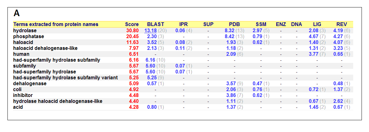
List of all matched protein name terms for 2gfh. The score in red is a measure of how strongly the term is predicted from the hits obtained by
the different methods. The scores in blue show each method’s contribution to the total score (with the number of relevant
sequences/structures shown in brackets in grey).(http://www.ebi.ac.uk/thornton-srv/databases/cgi-bin/pdbsum/)
- Higest score in GO - Hydrolase
- Followed by - Phosphatase
- Polymer: Haloacid dehalogenase-like hydrolase domain containing 4
Surface Properties
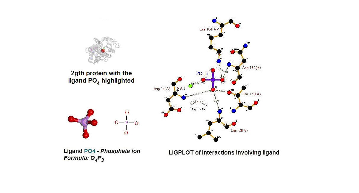 Figure 13. (A) Molecular structure of 2gfh with the ligand PO4. (B) Molecular and chemical structure of PO4. (C) Ligand interaction involving PO4. (Picture adapted from Profunc)
- Identify the likely biochemical function from the 3D structure
- Possible binding sites and potential ligands - PO4
- PO4 most likely be an active site and fuction
Phostphatase
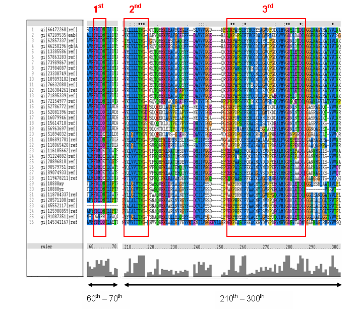
MSA of the 2gfh with 35 others proteins. Only the 60th – 70th and the 210th -300th amino acid
sequence were shown to illustrate the conserved and invariant regions. The 3 boxed-up sequences were either conserved or invariant regions.
- 2nd - threonine (T), asparagine (N) and glycine (G)
- 3rd - lysine (K) and aspartates (D)
- MSA corelate with with study done by Maliekal et al
- N-acetylneuraminic acid phosphatase orthologs shared 3 motifs found in phosphatases
- HAD (Haloacid dehalogenase-like) family
- Phosphatase activity: CO–P bond hydrolysis
- Dehalogenase activity: C–halogen bond hydrolysis
- Phosphonatase: C–P bond hydrolysis
- Phosphoglucomutase: CO–P bond hydrolysis and intramolecular phosphoryl transfer
Role in Human
- OMIM
- Haloacid dehalogenase-like hydrolase domain
- Gene map locus 20p11
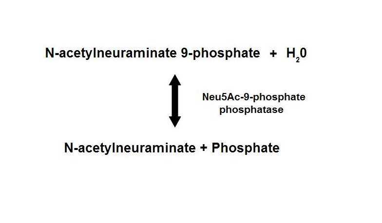
Dephosphorylation of Neu5Ac-9-P is a reversible reaction with an end product of Neu5Ac (sialic acid) and a free phosphate.
- Main form of sialic acid in vertebrates
- Important roles in protein-protein and cell-cell recognition
- Dependent on the presence of Mg2+
- Inhibited by vanadate and Ca2+
Role in Bacteria - E.coli
- Hydrolyze a wide range of phosphorylated metabolites
- Carbohydrates
- Nucleotides,
- Organic acids
- Kuznetsova et al - glycolysis and pentose phosphate pathway
- Fructose-1-phosphate
- Glucose-6-phosphate
- Mannose-6-phosphate
- 2-deoxyglucose-6-phosphate
- Fructose-6- phosphate
- Ribose-5-phosphate
- Erythrose- 4-phosphate
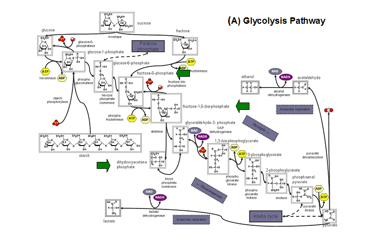
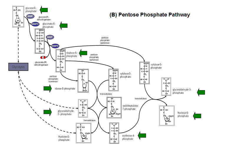
Schematic diagrams of glycolysis and pentose phosphate metabolic pathways. The green arrows show the substrates that are hydrolyzed by HADs
Press here to go Back


 Figure 13. (A) Molecular structure of 2gfh with the ligand PO4. (B) Molecular and chemical structure of PO4. (C) Ligand interaction involving PO4. (Picture adapted from Profunc)


