Locating The Bind Site For ACO In Coenzyme A Synthase: Difference between revisions
From MDWiki
Jump to navigationJump to search
No edit summary |
No edit summary |
||
| Line 1: | Line 1: | ||
ACO | ACO | ||
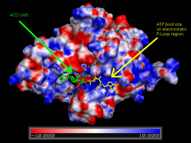
[[Image:COASYApbs_mapped_binding_cradle_shown_with_aligned_DPCK_IJJV_and_associated_ATP_ligand_biniding.jpg|framed|'''Figure 6 '''<BR> Surface map of ''Mus musculus'' Coenzyme A Synthase with electrostatic charge indicated, ATP ligand is included at P-Loop bind site of DPCK domain and ACO at the proposed Dephospho Coenzyme A Kinase cleft.|none]]<BR> | |||
{| cellpadding="0" | border="1" | |||
|+ '''Figure 13''' | |||
|- | |||
|- | |||
|[[Image:COASYClefts.jpg|thumb|400px|none]] | |||
|- | |||
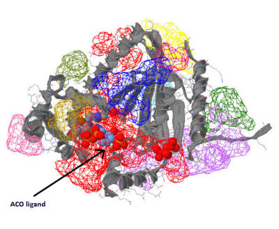
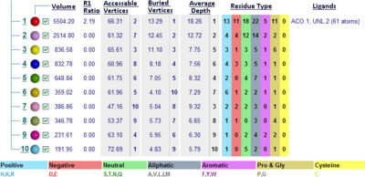
|[[Image:COASYCleft_table.jpg|thumb|400px|<BR> Location of binding clefts on Coenzyme A Synthase Mus. musculus with ACO and an unknown ligand (UNL).|none]] | |||
|- | |||
|} | |||
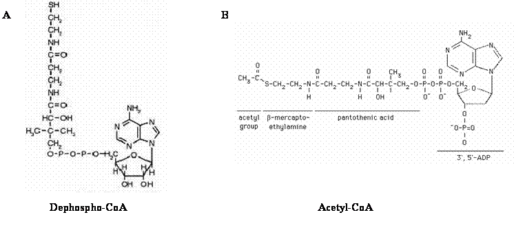
[[Image:ACO_and_DephosphoCoA_(fig_in_report).gif|framed|'''Figure 16'''<BR>Structures of Dephospho-CoA (A) and Acetyl-CoA (B). Structure of dephospho-CoA was reproduced from Daugherty et. al., 2002. Structure of Acetyl-CoA was reproduced from Baggot & Dennis, 1998.|none]]<BR> | |||
[[Future Directions COASY|Next??]] | [[Future Directions COASY|Next??]] | ||
[[Coenzyme A Synthase DPCK Domain Defining The P-Loop|Previous page]] | [[Coenzyme A Synthase DPCK Domain Defining The P-Loop|Previous page]] | ||
Revision as of 06:28, 11 June 2007
ACO


