Phytanoyl-CoA dioxygenase Structure: Difference between revisions
No edit summary |
No edit summary |
||
| Line 19: | Line 19: | ||
<br>There are high levels of steric hinderance if the approaching ligand comes from behind the molecule (<b>figure 1, figure 3</b>). | <br>There are high levels of steric hinderance if the approaching ligand comes from behind the molecule (<b>figure 1, figure 3</b>). | ||
There is a large hole in the structure of our protein (<b>figure 2, figure 4</b>). As steric hinderance is low and the surface area is maximized at this position, it would seem that this area should encompass the binding sites of our protein, or at least be the location of some form of interaction. The fact such a definitive hole in the structure of the protein exists lead us to investigate further properties around this region. | There is a large hole in the structure of our protein (<b>figure 2, figure 4</b>). As steric hinderance is low and the surface area is maximized at this position, it would seem that this area should encompass the binding sites of our protein, or at least be the location of some form of interaction. The fact such a definitive hole in the structure of the protein exists lead us to investigate further properties around this region. Liam will go into more details as to the function of the binding sites and a comparison to a similar protein in his section. | ||
[[Image:topology2.gif|center|framed|'''Figure 1'''<Br> Showing the topology of the protein. Reproduced from the European Bioinformatics Institute, <Br>http://www.ebi.ac.uk/thornton-srv/databases/pdbsum/2opw/domA01.gif]]<br> | [[Image:topology2.gif|center|framed|'''Figure 1'''<Br> Showing the topology of the protein. Reproduced from the European Bioinformatics Institute, <Br>http://www.ebi.ac.uk/thornton-srv/databases/pdbsum/2opw/domA01.gif]]<br> | ||
Revision as of 21:57, 9 June 2008
Originally, the structure of our protein was experimentally defined (by Zhang, Z., Butler, D., McDonough, M.A et.al.) via X-ray diffraction.
Name - Phytanoyl-CoA dioxygenase (PHYHD1)
Classification - Oxidoreductase
Resolution (Amstrongs) - 1.90
R-Value - 0.221 (obs, relatively low)
Space Group - P 3.1 2 1
Unit Cell Paramters (Amstrongs) - a = 91.97, b = 91.97, c = 81.61
Unit Cell Angles - alpha = 90.00, beta = 90.00, gamma = 120.00
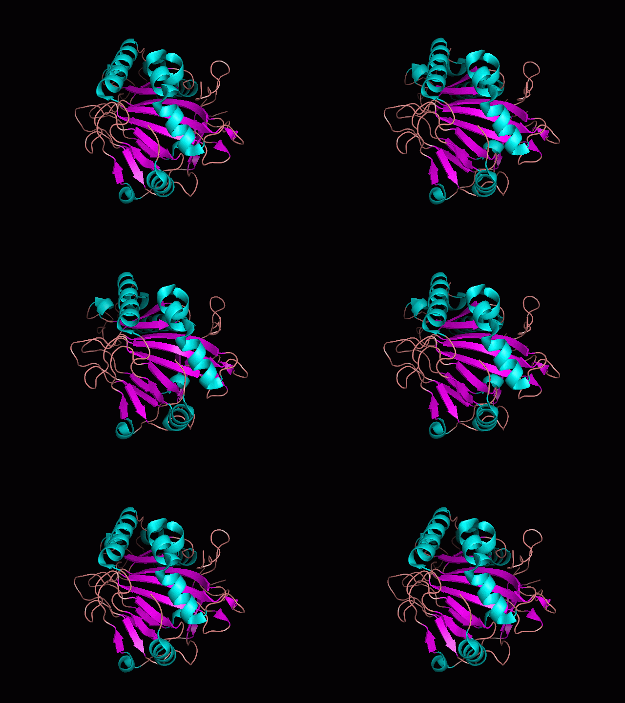
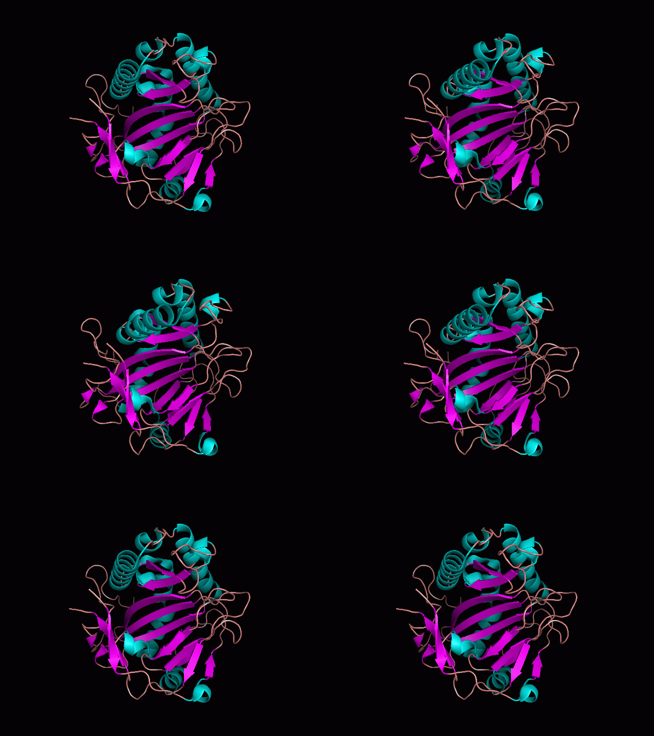
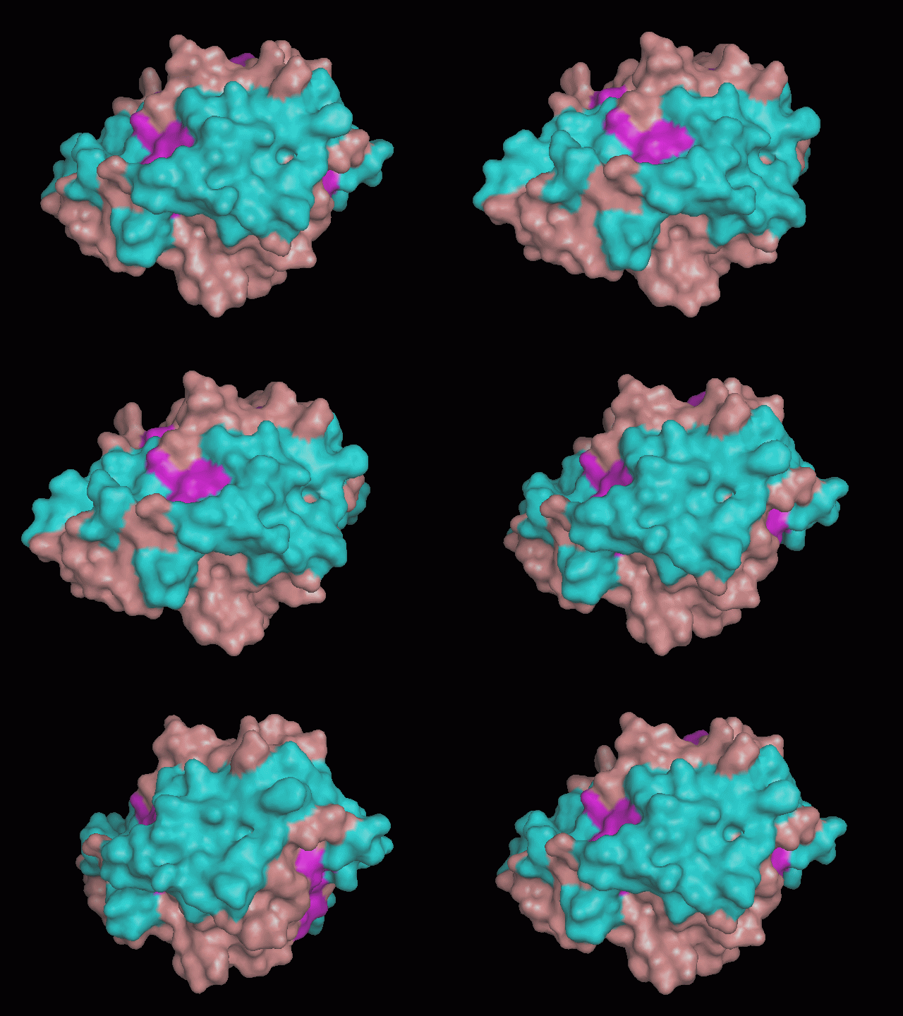
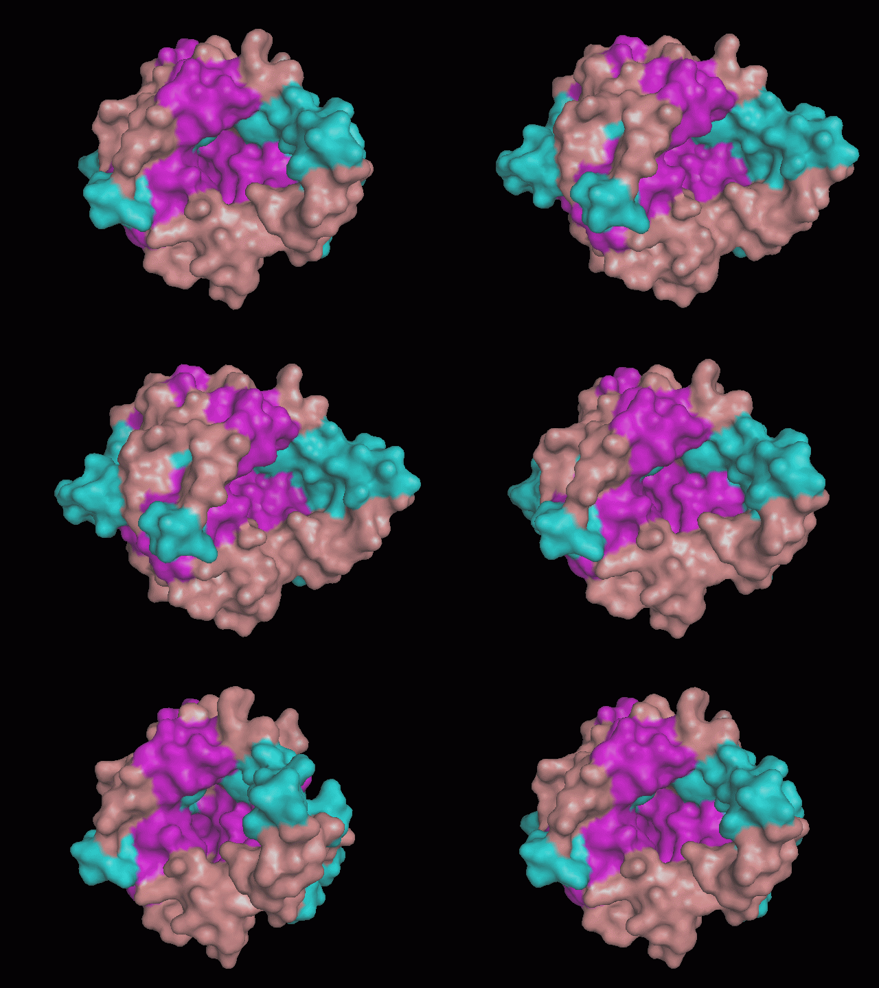
Using Pymol the image was visualized in a few different ways to get a general overview of the protein. The stick structures tended to be clutered so a surface rendering was done and a cartoon depiction. These are below.
There are high levels of steric hinderance if the approaching ligand comes from behind the molecule (figure 1, figure 3).
There is a large hole in the structure of our protein (figure 2, figure 4). As steric hinderance is low and the surface area is maximized at this position, it would seem that this area should encompass the binding sites of our protein, or at least be the location of some form of interaction. The fact such a definitive hole in the structure of the protein exists lead us to investigate further properties around this region. Liam will go into more details as to the function of the binding sites and a comparison to a similar protein in his section.
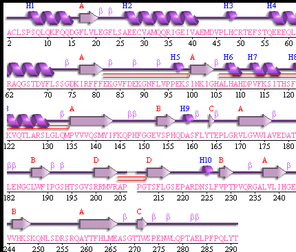
Showing the topology of the protein. Reproduced from the European Bioinformatics Institute,
http://www.ebi.ac.uk/thornton-srv/databases/pdbsum/2opw/domA01.gif
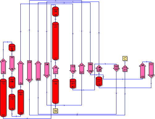
Showing another topology of the protein. Reproduced from the European Bioinformatics Institute,
http://www.ebi.ac.uk/
