Phytanoyl-CoA dioxygenase Structure
From MDWiki
Jump to navigationJump to search
File:Pocket.png
Figure 1
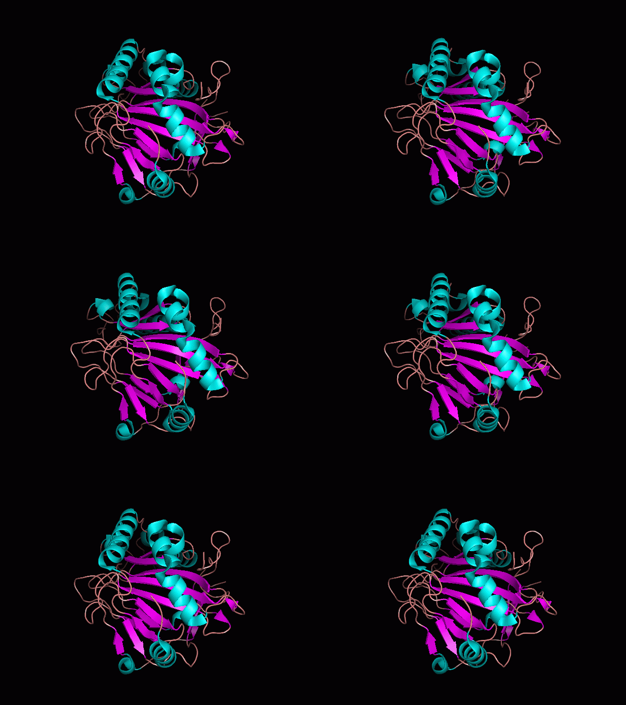
Using Pymol the protein was visualized. Structurally, the pocket can be seen in this picture, this is the area in which the ligand enters and binds.
Using Pymol the protein was visualized. Structurally, the pocket can be seen in this picture, this is the area in which the ligand enters and binds.

Figure 1
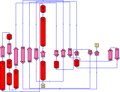
Showing the topology of the protein. Reproduced from the European Bioinformatics Institute,
http://www.ebi.ac.uk/thornton-srv/databases/pdbsum/2opw/domA01.gif
Showing the topology of the protein. Reproduced from the European Bioinformatics Institute,
http://www.ebi.ac.uk/thornton-srv/databases/pdbsum/2opw/domA01.gif

Figure 2
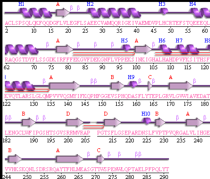
Showing another topology of the protein. Reproduced from the European Bioinformatics Institute,
http://www.ebi.ac.uk/
Showing another topology of the protein. Reproduced from the European Bioinformatics Institute,
http://www.ebi.ac.uk/
