Ribbon view of 2NXF: Difference between revisions
From MDWiki
Jump to navigationJump to search
No edit summary |
No edit summary |
||
| Line 2: | Line 2: | ||
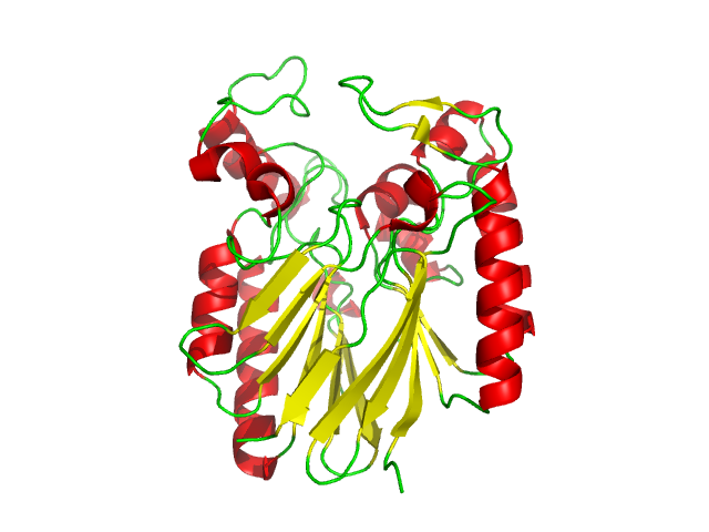
Ribbon view of 2NXF showing the 4-layer sandwich structure described by the SCOP classification. The structure shows that the outer layers of the protein are dominated by alpha | Ribbon view of 2NXF showing the 4-layer alpha/beta sandwich structure described by the SCOP classification. The structure shows that the outer layers of the protein are dominated by alpha helices, while the centre of the protein appears to be dominated by beta sheets. This structure contains 20 anti-parallel beta sheets, coloured yellow and 12 alpha helices, shown in red; coiled regions are shown in green. | ||
Revision as of 02:31, 30 May 2008
Ribbon view of 2NXF showing the 4-layer alpha/beta sandwich structure described by the SCOP classification. The structure shows that the outer layers of the protein are dominated by alpha helices, while the centre of the protein appears to be dominated by beta sheets. This structure contains 20 anti-parallel beta sheets, coloured yellow and 12 alpha helices, shown in red; coiled regions are shown in green.
