Uncategorized files
From MDWiki
Jump to navigationJump to search
Showing below up to 250 results in range #1,001 to #1,250.
 MsbA Petsko07.pdf 0 × 0; 144 KB
 MultipleBlastProtSeq.txt ; 9 KB
 Multipleestructuralalignmenta.jpeg 825 × 229; 54 KB
 Multiplestructuralalgnmentb.jpeg 893 × 214; 56 KB
 Multiseq align.jpg 839 × 331; 170 KB
 Mut4.gif 400 × 244; 10 KB
 Mutation1.png 220 × 77; 1 KB
 Mutation2.png 220 × 75; 1 KB
 Mutation3.png 240 × 75; 1 KB
 Mutation4.png 519 × 135; 5 KB
- N-terminus of LOC144557.txt ; 84 KB
 NKxD.jpg 146 × 653; 52 KB
 NKxD2.jpg 146 × 653; 52 KB
 NKxD3.jpg 468 × 742; 151 KB
 NKxD4.jpg 468 × 742; 151 KB
 NKxD5.jpg 514 × 736; 144 KB
 NUBP2BACK.jpg 200 × 200; 12 KB
 Names.jpg 1,051 × 437; 59 KB
 Nature encode.jpg 150 × 198; 32 KB
- Neighbortree.txt ; 100 KB
 Neighbourgene.jpg 770 × 680; 96 KB
- Neighbourtree2.txt ; 100 KB
 Nest.PNG 873 × 602; 79 KB
 Nest analysis result.PNG 545 × 320; 63 KB
 Nest analysis result.bmp 545 × 320; 511 KB
 Net.png 471 × 428; 29 KB
 Net image e 2H7YkMhiRnz6.png 471 × 456; 80 KB
 NewBOOT1000tree.png 934 × 720; 95 KB
 NewBOOTtree.png 1,050 × 812; 113 KB
 NewBOOTtree2.png 934 × 720; 95 KB
- Noble PLOSComputBiol2009.pdf 0 × 0; 130 KB
- NonredundantMSA.pdf 0 × 0; 454 KB
- NtermVS2oew.txt ; 208 KB
- NtermVs2cbi.txt ; 305 KB
 Nubp2front.jpg 200 × 200; 9 KB
 Nubp2top.jpg 200 × 200; 9 KB
 OG binding site.png 640 × 480; 26 KB
 Occurence.PNG 803 × 1,092; 57 KB
 Occurence.jpg 891 × 767; 48 KB
- Okamoto09.pdf 0 × 0; 305 KB
 Olf motif graph.GIF 623 × 382; 15 KB
 Olf motif table.GIF 915 × 994; 49 KB
 Olf trans pathway.gif 680 × 661; 39 KB
- Olfactory cGMP PDE2.PDF 0 × 0; 1.03 MB
 Ontology.jpg 608 × 468; 34 KB
 P1010632.jpg 1,620 × 2,236; 1.37 MB
 PBB GE DHRS1 213279 at fs.png 732 × 530; 12 KB
 PBB GE SELENBP1 214433 s at fs.png 732 × 530; 11 KB
 PDB.jpg 763 × 305; 41 KB
 PDB1.jpg 763 × 305; 41 KB
 PDBSUM.PNG 931 × 733; 145 KB
 PDBSUM.png 931 × 733; 145 KB
 PDBSum pblA.PNG 460 × 498; 31 KB
 PDB structure.jpg 250 × 250; 15 KB
 PDB sum 2.PNG 448 × 353; 38 KB
 PDBsum.JPG 440 × 652; 63 KB
 PDBsum.bmp 720 × 661; 1.36 MB
 PDBsum11.PNG 720 × 661; 90 KB
 PDBsum chain.JPG 878 × 734; 105 KB
 PDBsum cleft.PNG 803 × 681; 157 KB
 PDBsums.png 947 × 677; 69 KB
 PFam domains.png 638 × 477; 61 KB
 PHYH expression.png 520 × 790; 19 KB
 PICT1438.JPG 2,816 × 2,112; 1.11 MB
 PICT1438 copy.jpg 2,816 × 2,112; 1.11 MB
 PO4 400.gif 400 × 400; 1 KB
 PO4 interactions.png 389 × 406; 16 KB
 PPmotif.PNG 771 × 180; 27 KB
 PROFUNC Match To Existing PDB Stuctures.jpg 1,043 × 214; 45 KB
 PROFUNC Nest Analysis.jpg 645 × 363; 51 KB
 PROFUNC SSM.jpg 804 × 190; 43 KB
 PROKNOW-biological process.png 929 × 1,112; 20 KB
 PROKNOW2- molecular function.png 512 × 824; 9 KB
 PTS-1.jpg 282 × 66; 11 KB
 PTS-2.jpg 397 × 71; 15 KB
 PUBSUM.jpg 430 × 365; 18 KB
- Pastore febs07.pdf 0 × 0; 1.43 MB
 Pathway.jpg 773 × 261; 62 KB
 Pathways.png 421 × 590; 67 KB
- Pattern0.txt ; 31 bytes
 Pbio GOS 120.gif 120 × 120; 5 KB
 Pbl.png 640 × 480; 182 KB
 Pdb cartoon 2ece.png 680 × 846; 12 KB
 Pdb sum.png 864 × 648; 185 KB
 Pdbsum 1senA.gif 430 × 233; 11 KB
 Pdbsum 3fpu.gif 430 × 256; 13 KB
 Pdbsums archeal.PNG 894 × 528; 66 KB
 Pep 1.PNG 804 × 696; 91 KB
- Peptide Folding- When Simulation Meets Experiment.pdf 0 × 0; 270 KB
 Petri.jpg 388 × 385; 76 KB
 Pfam-a.jpg 1,066 × 108; 85 KB
 Pfam result.jpg 960 × 720; 114 KB
- Phillips04.pdf 0 × 0; 395 KB
 Phylo radial1.png 947 × 644; 30 KB
 Phylo radial1a.png 863 × 587; 42 KB
 Phylo rect1.png 1,007 × 517; 39 KB
 Phylo tree.jpg 1,146 × 633; 61 KB
 Phylogenetic Tree.jpg 1,280 × 1,024; 141 KB
 Phylogenic tree.JPG 680 × 563; 47 KB
 Phylogenic tree3.jpg 680 × 563; 47 KB
 Phylogram.png 1,280 × 768; 50 KB
 Pi helix.gif 58 × 20; 1 KB
 Picture1.jpg 793 × 655; 42 KB
 Picture2.jpg 333 × 437; 7 KB
 Picture3.jpg 412 × 395; 14 KB
 Picture4.png 1,465 × 941; 2.81 MB
 Picture5.jpg 1,502 × 811; 337 KB
 Picture6.jpg 1,427 × 704; 176 KB
 Picture 1.png 495 × 455; 180 KB
 Picture 2.png 514 × 407; 40 KB
 Plasm.png 939 × 461; 202 KB
 Plasmodium.png 939 × 461; 202 KB
 Platypus.png 1,261 × 629; 120 KB
 Po4 ligand.JPG 763 × 879; 72 KB
 Pocket.gif 640 × 480; 1.65 MB
 Pocket.jpg 1,062 × 570; 74 KB
 Pocket surface.gif 640 × 480; 3.36 MB
 Possiblecatalyticresidues.png 640 × 434; 92 KB
 Pprofunc superfamily.JPG 1,280 × 1,024; 127 KB
- Prestegard ms1.pdf 0 × 0; 36 KB
- Prestegard ms2.pdf 0 × 0; 67 KB
 Pretty.png 640 × 434; 79 KB
 Pretty protein.PNG 613 × 660; 387 KB
 ProFunc Image.jpg 480 × 480; 19 KB
 ProFunc ImageJen.jpg 480 × 480; 19 KB
 Profunc, GO.png 960 × 720; 226 KB
 Profunc.jpg 775 × 582; 74 KB
 Profunc2.jpg 1,089 × 261; 73 KB
 ProfuncNest2.PNG 651 × 577; 41 KB
 ProfuncNests.JPG 651 × 614; 235 KB
 Profunc GO.jpg 960 × 720; 102 KB
- Profunc NAR2005.pdf 0 × 0; 1.95 MB
- Profunc jmb2007.pdf 0 × 0; 908 KB
 Profunc nest.PNG 622 × 195; 13 KB
 Profunc nest.bmp 622 × 195; 356 KB
 Profunc nests.JPG 651 × 614; 84 KB
 Profunc nests.jpg 651 × 614; 84 KB
- Proknow Eisenberg Structure2005.pdf 0 × 0; 480 KB
 Prositeprediction.jpg 477 × 599; 23 KB
 Prositescan.png 477 × 599; 23 KB
- Protdist1.txt ; 1.24 MB
- Protdist2.txt ; 1.24 MB
- Protein-result.txt ; 6 KB
- Protein-results.txt ; 6 KB
 Protein2.JPG 2,102 × 434; 79 KB
 Protein interaction.jpg 471 × 456; 80 KB
 Protein sequence.JPG 606 × 146; 33 KB
 Protein showing zinc pymol.png 1,060 × 906; 232 KB
 PsaA docked to E-cadherin in simulation cell.png 1,044 × 941; 739 KB
- Psiblast.txt ; 1.03 MB
 PyMOL 2oycA vs 2cfsA.jpg 555 × 397; 32 KB
- Error creating thumbnail: convert: Expected 8192 bytes; found 8119 bytes `/var/www/mediawiki/upload/2/29/Pymol.PNG.png' @ warning/png.c/MagickPNGWarningHandler/1669. convert: Read Exception `/var/www/mediawiki/upload/2/29/Pymol.PNG.png' @ error/png.c/MagickPNGErrorHandler/1643. convert: no images defined `/tmp/transform_ee25e6271e32.png' @ error/convert.c/ConvertImageCommand/3229. Error code: 1Pymol.PNG.png 1,060 × 906; 713 KB
 Pymol.png 640 × 434; 167 KB
 Pymol hsp movie0001.png 580 × 482; 53 KB
 Pymol ss1.PNG 406 × 370; 59 KB
- Pyridoxal Phosphatase MSA sequences.pdf 0 × 0; 694 KB
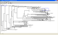
 Pyridoxal Phosphatase Rectangular Tree.jpg 3,508 × 2,480; 485 KB
- Pyridoxal phosphatase BLAST.pdf 0 × 0; 250 KB
 Pyridoxal phosphatase MSA Page 1.jpg 783 × 377; 59 KB
 Pyridoxal phosphatase MSA Page 2.jpg 783 × 377; 170 KB
 Pyridoxal phosphatase MSA Page 3.jpg 783 × 377; 202 KB
 Pyridoxal phosphatase MSA Page 4.jpg 306 × 369; 73 KB
 Pyridoxal phosphatase MSA sequences conserved .jpg 927 × 928; 457 KB
 Pyridoxal phosphatase Radial Cladogram.jpg 3,508 × 2,480; 477 KB
- Pyridoxal phosphatase Rectangular Phylogeny.pdf 0 × 0; 176 KB
 Pyridoxal phosphatase Rectangular Tree discussion1.jpg 1,768 × 704; 97 KB
 Pyridoxal phosphatase conserved image.jpg 640 × 480; 105 KB
- Querol2003.pdf 0 × 0; 827 KB
- Querol2006.pdf 0 × 0; 145 KB
 Question mark.jpg 10 × 18; 539 bytes
 RNA template 1hu3.gif 732 × 377; 33 KB
 RR1.png 798 × 577; 574 KB
 RR2.png 791 × 521; 506 KB
 RR3.png 793 × 526; 451 KB
 Rad.JPG 967 × 839; 71 KB
 Rad2.JPG 1,048 × 859; 67 KB
 Radial.JPG 967 × 839; 71 KB
 Radial2.JPG 1,048 × 859; 67 KB
 Radialtree.JPG 656 × 607; 62 KB
 Radiationtreexx22.jpg 789 × 722; 38 KB
 Ramachandran.jpg 786 × 393; 47 KB
 Ramachandran 1.jpg 816 × 391; 34 KB
 Rat fatty acid binding protein 2IFB.jpg 500 × 500; 47 KB
 Rbsfunction.jpg 662 × 201; 31 KB
- Rdc key equations.pdf 0 × 0; 271 KB
 Reaction.jpg 506 × 146; 10 KB
 Reaction.png 866 × 783; 20 KB
 Reaction1.png 530 × 242; 10 KB
 ReactionASK.png 416 × 92; 13 KB
 Reaction Arrow.jpg 26 × 21; 775 bytes
 Red x.png 25 × 28; 3 KB
 Redox Halves.png 892 × 227; 7 KB
 Region1.png 1,280 × 1,024; 972 KB
 Region2.png 1,272 × 929; 153 KB
 Region3.png 1,272 × 929; 138 KB
 Rhodopseudomonas palustris chromosome.jpg 900 × 628; 88 KB
 Ribbon structure.png 640 × 434; 82 KB
 Rootedtree1.jpg 1,170 × 618; 53 KB
 Rootedtreeboot.jpg 1,170 × 618; 50 KB
 Rootedtreecol1.jpg 1,170 × 618; 67 KB
 Rootedtreecol2.jpg 1,170 × 618; 71 KB
- Roux1999 BiophysChem78-1.pdf 0 × 0; 211 KB
- Ruscio2008 PNAS105-9204.pdf 0 × 0; 697 KB
- Russel Structure2004.pdf 0 × 0; 287 KB
 SABLE.gif 550 × 664; 19 KB
 SAMPL.png 522 × 132; 49 KB
 SAS Aligned Sequences.jpg 1,190 × 243; 66 KB
 SBP with Sele Atom.png 640 × 480; 161 KB
 SBP with Sele atom.png 640 × 480; 161 KB
 SBP with sele atom.png 640 × 480; 161 KB
 SCOP.JPG 1,167 × 122; 36 KB
 SECONDARY.jpg 503 × 471; 9 KB
- SPLIT combined alan.pdf 0 × 0; 34.95 MB
 SSAME.png 640 × 434; 97 KB
 SSM1.PNG 791 × 520; 50 KB
 SSM2.PNG 792 × 402; 54 KB
 SSMPhosphatases.jpg 3,124 × 1,013; 551 KB
- SSMPhosphatases.pdf 0 × 0; 21 KB
 SSP.png 1,216 × 445; 49 KB
 STSsite1.png 660 × 629; 190 KB
 STSsite2.png 660 × 629; 190 KB
 STSwhole.png 616 × 603; 215 KB
 SURFACE.jpg 986 × 768; 118 KB
 Schematic.jpg 590 × 444; 35 KB
- Schultz chembiochem02.pdf 0 × 0; 101 KB
- Schultz currentopinionChemBiol05.pdf 0 × 0; 401 KB
- Schultz nature methods07.pdf 0 × 0; 389 KB
- Schultz pnas02.pdf 0 × 0; 562 KB
 Scop.png 404 × 217; 44 KB
 Secondary.jpg 925 × 661; 92 KB
 Secondary Structure.png 537 × 623; 42 KB
 Secondary Structure 2cfsA.jpg 486 × 765; 77 KB
 Secondary Structure 2oycA.jpg 466 × 731; 69 KB
 Secondary structure.gif 430 × 600; 24 KB
 Secondary structure.jpg 540 × 390; 23 KB
 Secondary structure.png 540 × 390; 23 KB
- Secondary structure.txt ; 559 bytes
 Secondary structure comparsion.jpg 1,215 × 480; 70 KB
- Secondary structures.pdf 0 × 0; 13.24 MB
 SelNonredundantMSA.PNG 1,344 × 646; 113 KB
 SelNonredundantMSA.jpg 2,120 × 1,590; 1.45 MB
- SelNonredundantMSA.pdf 0 × 0; 171 KB
- Seminario Hess2FF.pdf 0 × 0; 480 KB
 Seq1.jpg 732 × 78; 4 KB
 Seq2.jpg 593 × 111; 14 KB
 Sequence.jpg 791 × 432; 36 KB
 Sequence1-cml.jpg 750 × 398; 102 KB
 Sequence & secondary structure PDB.bmp 0 × 0; 906 KB


