Uncategorized files
From MDWiki
Jump to navigationJump to search
Showing below up to 250 results in range #501 to #750.
 Cleft Analysis 2cfsA.jpg 911 × 696; 85 KB
 Cleft Analysis 2oycA.jpg 905 × 720; 88 KB
 Cleft Analysis PyMOL 2cfsA V1.jpg 494 × 404; 42 KB
 Cleft Analysis PyMOL 2cfsA V2.jpg 433 × 429; 44 KB
 Cleft Analysis PyMOL 2oycA V1.jpg 456 × 381; 41 KB
 Cleft Analysis PyMOL 2oycA V2.jpg 463 × 403; 42 KB
 Cleft analysis 2i2O.jpg 0 × 0; 1.1 MB
 Clefts.JPG 1,059 × 850; 132 KB
 Clus.jpg 389 × 235; 20 KB
 Clustal.jpg 394 × 329; 21 KB
 Clustal1.jpg 1,282 × 377; 182 KB
 Clustal2.jpg 1,278 × 375; 194 KB
 Clustal3.jpg 1,276 × 387; 206 KB
 ClustalX-alignment.JPG 1,030 × 837; 336 KB
 ClustalX.jpg 1,203 × 618; 381 KB
 ClustalX image.JPG 1,203 × 618; 390 KB
 ClustalX omit.jpg 1,158 × 617; 339 KB
 Clustal 0-130.jpg 972 × 481; 113 KB
 Clustal 130-260.jpg 875 × 432; 124 KB
 Clustal 260-390.jpg 875 × 432; 131 KB
 Clustal 390-520.jpg 876 × 434; 133 KB
 Clustal 520-650.jpg 875 × 432; 110 KB
 Clustal Alignment top 5.txt ; 4 KB
 Clustal X.jpg 1,203 × 618; 381 KB
 Clustal results.jpg 970 × 483; 142 KB
 Clustalexample.JPG 366 × 270; 41 KB
 Clustalexample.bmp 366 × 270; 41 KB
 Clustalx.pdf 0 × 0; 1.24 MB
 Clustalx.png 859 × 1,320; 140 KB
 Clustalx1.JPG 838 × 572; 189 KB
- Clustalx1.jpg 0 × 0; 3.75 MB
 Clustalx1a.jpg 1,280 × 1,024; 438 KB
 Clustalx2.JPG 838 × 571; 208 KB
 Clustalx2.jpg 1,280 × 1,024; 475 KB
 Clustalx3.JPG 838 × 582; 218 KB
 Clustalx3a.jpg 1,280 × 1,024; 503 KB
 Clustalx4a.jpg 1,280 × 1,024; 444 KB
 Clustalx5a.jpg 1,280 × 1,024; 445 KB
 Clustalx6a.jpg 1,280 × 1,024; 482 KB
 Clustalx taken from residue 98-350.png 1,280 × 771; 118 KB
 Clustalx taken from residue 98-350 Part2.png 1,280 × 769; 112 KB
 Cluster.png 640 × 434; 143 KB
- Clustering methods.pdf 0 × 0; 1.88 MB
 Clutsalw1.JPG 941 × 831; 132 KB
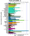 CoAA - Pantothenate kinase expression data (mouse).png 520 × 610; 14 KB
 CoAD reaction.gif 977 × 189; 5 KB
 CoAE reaction.gif 1,112 × 197; 5 KB
 CoA Synthesis localistion.png 520 × 610; 14 KB
 CoA Synthesis pathway.gif 454 × 372; 4 KB
 CoA structure.png 300 × 117; 25 KB
 CoA structure 2.png 300 × 126; 5 KB
 CoA synthase localisation.png 520 × 610; 14 KB
 CoAsy.jpg 700 × 700; 27 KB
 Coasy2f6rapredictedligandbindingsites.JPG 640 × 512; 31 KB
 CoasyCphmodelpredictedligandbindingsites.JPG 640 × 512; 33 KB
 CoasyPocketaco.JPG 1,136 × 756; 108 KB
 CoasyPocketunl.JPG 1,111 × 757; 111 KB
 Collaboration ven diagram.PNG 535 × 469; 11 KB
 Compar1.png 640 × 408; 89 KB
 Compar2.png 640 × 408; 93 KB
 Comparison 2ijz and 1vhe.JPG 942 × 299; 51 KB
 Comparison 2ijz and 1vhe sequence.JPG 576 × 627; 107 KB
 Comparison 2ijz and 1vhe sequence2.JPG 576 × 627; 107 KB
 Comparison 2ijz and 1xfo3.JPG 817 × 334; 52 KB
 Comparison 2ijz and 2cf4.JPG 1,066 × 312; 60 KB
 Comparison stuff (note ligands).jpeg 1,035 × 631; 133 KB
 Comparison stuff 2.jpeg 971 × 625; 99 KB
 Confidence.jpeg 1,093 × 288; 39 KB
 Confidence.png 533 × 473; 52 KB
 Confidence interaction with names.PNG 727 × 540; 72 KB
 Confidence interaction with names.png 727 × 540; 72 KB
 Conformation type of ERp18.png 592 × 425; 99 KB
- Connolly1983 JApplCrystallogr16-548.pdf 0 × 0; 1.49 MB
 Conres.jpg 757 × 595; 130 KB
 Conres.png 527 × 645; 123 KB
 Consense.jpg 1,887 × 1,187; 134 KB
- Consense.txt ; 33 KB
 Consensetree.jpg 1,887 × 1,187; 80 KB
 Conserved.JPG 593 × 819; 40 KB
 Conserved.jpg 512 × 384; 316 KB
- ConservedRegion 1.bmp 0 × 0; 4.47 MB
 ConservedRegion 1.png 1,024 × 687; 95 KB
 ConservedRegion 2.png 1,021 × 681; 89 KB
 ConservedRegion 3.png 1,024 × 685; 80 KB
 Conserved regions.JPG 420 × 475; 24 KB
 Conserved regions.jpg 640 × 480; 471 KB
 Conserved regions of 2ijz-A.JPG 422 × 401; 26 KB
 Conservedtext.JPG 420 × 518; 28 KB
- Convergence of sampling in protein simulations.pdf 0 × 0; 410 KB
 Cross.jpg 25 × 28; 1 KB
 Crystal Structure 2cfsA.jpg 240 × 226; 10 KB
 DALI2pbl.png 725 × 144; 9 KB
 DALI 2cfsA Updated.jpg 796 × 452; 116 KB
- DALI RESULT.txt ; 1.51 MB
 DAL HSLs.png 730 × 172; 12 KB
 DSCF1385.JPG 2,848 × 2,136; 1.59 MB
 DSCF1386.JPG 2,136 × 2,848; 1.49 MB
 DSCF1387.JPG 2,848 × 2,136; 1.57 MB
 DSC 0127.jpeg 276 × 354; 85 KB
 DSC 0127.jpg 276 × 354; 85 KB
 Dali.PNG 925 × 724; 76 KB
 Dali.bmp 925 × 724; 1.92 MB
 Dali.jpg 1,008 × 247; 56 KB
 Dali33.PNG 760 × 603; 63 KB
 Dali Results.JPG 684 × 616; 142 KB
 Dali cutoff.jpg 976 × 287; 69 KB
 Dali output.jpg 721 × 345; 166 KB
 Dali output2.jpg 721 × 345; 89 KB
 Dali output3.jpg 721 × 345; 91 KB
 Dali results.png 653 × 333; 35 KB
 Dalii.PNG 760 × 603; 63 KB
 DanielTopology.gif 502 × 384; 9 KB
 DanielTopology2.gif 430 × 367; 19 KB
- Davis AngewChemIntEd03.pdf 0 × 0; 682 KB
- Davis DrugDiscovToday08.pdf 0 × 0; 1.52 MB
 Dehydrogenasereductase (SDR family) member 1 phylogram.png 835 × 608; 51 KB
- DesaltTrypticPeptide.pdf 0 × 0; 45 KB
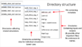 Directory structure 1.png 1,000 × 563; 152 KB
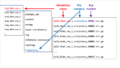 Directory structure 2.png 1,000 × 563; 142 KB
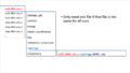 Directory structure 3.png 1,000 × 564; 77 KB
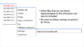 Directory structure 4.png 1,000 × 563; 95 KB
- Distance Matrix Calcuation.txt ; 16 KB
 Document15 07.png 584 × 819; 27 KB
 Document17 01.png 1,189 × 426; 39 KB
 Document17 03.png 1,210 × 514; 45 KB
 Document18 01.png 807 × 160; 4 KB
 Document19 08.png 616 × 438; 59 KB
 Document20 01.png 740 × 509; 147 KB
 Document20 02.png 815 × 543; 97 KB
 Document2 01.png 784 × 458; 91 KB
 Document2 04.png 744 × 865; 666 KB
 Document2 05.png 229 × 145; 3 KB
 Document5 01.png 556 × 354; 7 KB
 Document6 01.png 1,214 × 652; 28 KB
 Document6 02.png 1,214 × 652; 29 KB
 Document7 01.png 1,214 × 652; 29 KB
 Document7 02.png 1,214 × 652; 75 KB
 Document7 03.png 1,214 × 682; 182 KB
 Document7 04.png 1,104 × 620; 50 KB
 Document7 05.png 1,214 × 652; 314 KB
 Document7 07.png 1,214 × 652; 94 KB
 Document7 11.png 1,214 × 652; 55 KB
 Document9 01.png 720 × 617; 489 KB
 Domain--1.jpg 361 × 345; 33 KB
 Domain--2.jpg 353 × 364; 50 KB
 Domain-1.jpg 510 × 510; 61 KB
 Domain.JPG 395 × 104; 7 KB
 Domain.jpg 804 × 696; 93 KB
 Domain1.jpg 357 × 175; 40 KB
 Domain111.jpg 640 × 400; 12 KB
 Domain2.JPG 395 × 104; 7 KB
 Domain arrangement.jpg 493 × 212; 88 KB
 Domain pic.jpg 448 × 304; 71 KB
 Domain pic2.jpg 512 × 384; 112 KB
 Domain pic3.jpg 512 × 384; 153 KB
 Domains.jpg 804 × 696; 53 KB
 Domains 2F6r.jpg 623 × 480; 30 KB
 Dotlet.PNG 508 × 336; 85 KB
 Doxorubicin bound surface POPC CLR bilayer.png 719 × 721; 1 MB
 DrosophilaVsHuman.png 600 × 300; 5 KB
- Drug Follow The Rules.pdf 0 × 0; 577 KB
 EIF4G w groove.JPG 512 × 384; 9 KB
 EIF4G w groove2.JPG 186 × 263; 7 KB
- EMI.jpg 0 × 0; 879 KB
 EMIi.jpg 343 × 336; 26 KB
 EPOR-EMP33 asu.png 1,181 × 816; 1.02 MB
 Edo4 ligand.JPG 762 × 783; 64 KB
 Electrostatic.gif 640 × 434; 4.14 MB
 Electrostatic potential.png 507 × 481; 229 KB
 Electrostatic potential (molecular surface).png 507 × 481; 229 KB
 Eskimos.jpg 104 × 104; 4 KB
 Esp.jpg 397 × 515; 24 KB
 Example.jpg 596 × 591; 78 KB
- Example1.jpg ; 131 KB
 Examplec.jpg 470 × 359; 25 KB
 Examplex.jpg 1,006 × 179; 30 KB
 Exp.jpg 520 × 790; 23 KB
 Export.png 1,463 × 848; 40 KB
 Expression.png 520 × 610; 13 KB
 Expression PHYHD1.png 520 × 790; 18 KB
 Expressionlev.jpg 640 × 724; 59 KB
 Expressionlev2.jpg 640 × 724; 63 KB
 External unifi order.png 987 × 796; 128 KB
- ExtractIDs.doc ; 302 KB
 FASTA.jpg 890 × 400; 94 KB
- FASTA.txt ; 249 bytes
 FASTA2.jpg 890 × 400; 99 KB
- FASTA2.txt ; 262 bytes
- FASTA fascin1 all.txt ; 56 KB
 Facscin broad bootstrap.jpg 802 × 588; 60 KB
- Fascin1 fasta.txt ; 1 KB
 Fascin CASTp back.PNG 936 × 1,050; 133 KB
 Fascin CASTp front.PNG 940 × 1,050; 132 KB
 Fascin bootstrap 500rep.jpg 1,159 × 852; 78 KB
 Fascin bootstrap animalia.insecta.jpg 933 × 845; 84 KB
 Fascin bootstrap animaliainsecta.jpg 933 × 845; 84 KB
 Fascin dali.PNG 925 × 724; 76 KB
 Fascin domains and conserved.png 640 × 434; 179 KB
 Fascin domains and conserved back.png 640 × 434; 178 KB
 Fascin electro back binding.PNG 640 × 480; 295 KB
 Fascin electro front binding.PNG 640 × 480; 317 KB
 Fascin movie.gif 640 × 434; 3 MB
 Fascin multiple alignment.jpg 1,184 × 562; 225 KB
 Fascin sec struc and domain.PNG 792 × 1,553; 116 KB
 Fascin secondary structure.png 640 × 480; 113 KB
- Fasta seq output of entrez.txt ; 31 KB
- Fewerseqforalign.txt ; 7 KB
 Fig1 chains.jpg 707 × 571; 66 KB
 Fig1 properties.JPG 962 × 1,112; 176 KB
 Fig1 properties.jpg 1,246 × 1,134; 233 KB
 Fig2.JPG 274 × 491; 24 KB
 Fig2 properties.jpg 960 × 535; 110 KB
 Fig3.JPG 445 × 757; 60 KB
 Fig3 Mg.jpg 1,042 × 491; 66 KB
 Fig4.JPG 415 × 939; 78 KB
 Fig4 class&fold.jpg 469 × 272; 21 KB
 Fig5 MSA.jpg 958 × 960; 186 KB
 Fig6 residues.jpg 580 × 489; 33 KB
- Fig 1.bmp 0 × 0; 1.96 MB
 Fig 2.PNG 556 × 354; 7 KB
 Figtree.PNG 1,024 × 768; 257 KB
 Figure 1.png 561 × 549; 9 KB
 Figure 10.png 799 × 313; 91 KB
 Figure 11.png 803 × 419; 88 KB
 Figure 12.png 759 × 511; 116 KB
 Figure 13.png 762 × 600; 155 KB
 Figure 14.png 795 × 646; 128 KB
 Figure 15.png 758 × 640; 120 KB
 Figure 16.png 755 × 642; 127 KB
 Figure 17.png 403 × 87; 10 KB
 Figure 18.png 720 × 270; 62 KB
 Figure 19.png 1,211 × 713; 199 KB
 Figure 2.png 966 × 589; 146 KB
 Figure 3.png 338 × 596; 77 KB
 Figure 4.png 762 × 539; 107 KB
 Figure 5.png 756 × 579; 84 KB
 Figure 6.png 755 × 625; 83 KB
 Figure 7.png 755 × 743; 83 KB
 Figure 8.png 753 × 321; 58 KB
- File.jpg 0 × 0; 4.47 MB
 Final CoA synthesis pathway 2.gif 929 × 257; 73 KB
 Final fish eye tree.png 799 × 618; 93 KB
 First draft.JPG 652 × 917; 82 KB
 Flascin 1.jpg 596 × 591; 78 KB


.png)
.png)
.png)


.jpeg)


_member_1_phylogram.png)


.png)


