Uncategorized files
From MDWiki
Jump to navigationJump to search
Showing below up to 250 results in range #751 to #1,000.
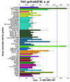
 Flj32549 expression profiles.GIF 1,110 × 1,458; 74 KB
 Fold.bmp 0 × 0; 339 KB
 Fold.jpg 640 × 181; 58 KB
 FoldMatch.jpg 793 × 512; 85 KB
 Fold analysis.jpg 733 × 186; 70 KB
 Frishman Proteins95.pdf 0 × 0; 1.32 MB
 Front view.png 640 × 408; 157 KB
 Fuc.jpg 324 × 178; 7 KB
 Fugue.png 896 × 717; 175 KB
 Function.jpg 651 × 400; 54 KB
 GGG and SYK.png 640 × 434; 148 KB
 GNP.jpg 800 × 300; 7 KB
 GNP1.jpg 800 × 300; 7 KB
 GO.png 786 × 187; 65 KB
 GO1.JPG 936 × 400; 68 KB
 GO ontology terms 2i2O.gif 144 × 336; 3 KB
 GOterm.JPG 962 × 748; 77 KB
- GPCR-A xray rev09.pdf 0 × 0; 636 KB
- GPCR srt 1 day ProtSci09.pdf 0 × 0; 495 KB
- GROMOS tutorial.pdf 0 × 0; 569 KB
 Galactose oxidase.png 576 × 396; 103 KB
 Gen posre.txt ; 2 KB
 Gene Ontology.bmp 380 × 86; 96 KB
 Gene ontology.PNG 755 × 400; 11 KB
 Geneatlas human.png 520 × 790; 19 KB
 Geneatlas mouse.png 520 × 610; 13 KB
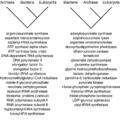 Geneologie2.png 667 × 665; 64 KB
 Geneprod.gif 555 × 88; 2 KB
 General structure of protein.png 640 × 480; 162 KB
 Genomic.gif 600 × 63; 931 bytes
 Genomic.jpg 812 × 355; 86 KB
 Getimg.gif 435 × 393; 23 KB
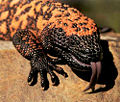 Gila.jpg 500 × 424; 61 KB
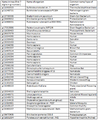 Gitable.png 463 × 567; 35 KB
- Golding pascual jmr82.pdf 0 × 0; 685 KB
- Golding stubbs RoyalSoc.pdf 0 × 0; 738 KB
- Golding stubbs jmr79.pdf 0 × 0; 886 KB
- Golding stubbs jmr80.pdf 0 × 0; 793 KB
 Green tick.png 24 × 24; 2 KB
- Gromos53a5a6.pdf 0 × 0; 181 KB
- Gromos sugar Hünenberger.pdf 0 × 0; 337 KB
- Gross Science2009.pdf.pdf 0 × 0; 972 KB
- Grottesi2002.pdf 0 × 0; 246 KB
- Group Tasks.doc ; 46 KB
- Group tasks.doc ; 46 KB
- Guanidination.pdf 0 × 0; 83 KB
- Guillot JMolLiq2002.pdf 0 × 0; 3.41 MB
 Gurad final.JPG 718 × 285; 30 KB
 Gurad final.bmp 718 × 285; 600 KB
- Guvench PLOSVomputBiol2009.pdf 0 × 0; 427 KB
- H-bond.jpg 601 × 466; 67 KB
 HDHD2 and predicted functional partner.png 464 × 791; 8 KB
 HDHD2 at.png 520 × 790; 19 KB
 HMM superfamily.JPG 822 × 816; 75 KB
 HSL alignment.png 621 × 600; 397 KB
 HSL thermophillic alignment.png 680 × 617; 60 KB
- Hammes-Schiffer AccChemRes2009.pdf 0 × 0; 318 KB
- Hansmann93.pdf 0 × 0; 1.05 MB
- Hansmann96.pdf 0 × 0; 568 KB
 Hbonding.png 640 × 434; 56 KB
- He5437.pdf 0 × 0; 473 KB
 Hessian.png 393 × 252; 5 KB
- HomoBLAST.aln ; 2.93 MB
- HomoCLUSTAL.aln ; 131 KB
 Horiike.PNG 348 × 346; 77 KB
 Human-3fdf.jpeg 502 × 483; 33 KB
- HumanOrthologProtSeq.txt ; 534 bytes
- HumanOrthologProtSeqBlast1.txt ; 52 KB
 HumanSymatlas.png 520 × 790; 18 KB
 Human Ssu72 BLAST result.PNG 652 × 367; 30 KB
 Human Ssu72 BLAST result.bmp 652 × 367; 701 KB
 Human geneAtlas.png 520 × 790; 19 KB
- Human homologs clean.fasta.txt ; 11 KB
 Humanhomolog.jpeg 692 × 508; 31 KB
 Humanproteins-cml.jpg 750 × 275; 18 KB
 Hydrolase.png 807 × 160; 4 KB
- Hydrophillic.jpg 601 × 466; 71 KB
- Hydrophobic.jpg 601 × 466; 77 KB
 Hydrophobic.png 582 × 411; 82 KB
 HydrophobicDHRS1.png 640 × 434; 113 KB
 Hydrophobicity.png 888 × 637; 100 KB
 Hydrophobicity.png.jpeg 592 × 425; 40 KB
 Hydrophobicity 2.png 527 × 434; 101 KB
 Hydrophobicity plot.PNG 605 × 408; 14 KB
 Hypotheticalmsa1xx.jpg 1,190 × 920; 214 KB
 Hypotreexx21.jpg 1,190 × 920; 45 KB
 IGP5020.jpg 600 × 401; 69 KB
 IGP5021.jpg 600 × 401; 55 KB
 IMG.jpg 1,613 × 1,063; 291 KB
 IMG 2.jpg 415 × 336; 22 KB
 INTER.jpg 593 × 333; 34 KB
 INTERPRO.jpg 593 × 333; 34 KB
 Icon-power-button.gif 28 × 28; 310 bytes
- Ids.txt ; 733 bytes
- Ids2.txt ; 879 bytes
 IfuA-structure.png 1,144 × 344; 241 KB
 Image-Dotlet.PNG 508 × 336; 85 KB
 Image001.png 1,257 × 529; 144 KB
 Image002.jpg 415 × 175; 18 KB
 Image003.png 1,260 × 544; 127 KB
 Image005.png 1,260 × 612; 172 KB
 Image007.png 609 × 109; 5 KB
 Image009.png 427 × 608; 55 KB
 Image011.png 421 × 611; 55 KB
 Image021.jpg 1,280 × 960; 221 KB
 Image021.png 671 × 493; 45 KB
 Image022.jpg 1,280 × 960; 199 KB
 Image027.jpg 1,280 × 960; 261 KB
 Image029.jpg 1,280 × 960; 285 KB
 Image 1.png 640 × 434; 134 KB
 Image 2.png 640 × 434; 136 KB
- Error creating thumbnail: File missingImage PROFUNC.jpg 384 × 384; 26 KB
 Image of CC bvbresidues.png 640 × 434; 258 KB
 Image of CC residues.png 640 × 434; 258 KB
 Image of ERp16.png 640 × 434; 139 KB
 Image of ERp16 only active site no ions.png 640 × 434; 138 KB
 Image of ERp18 conserved secondary structure.png 640 × 434; 82 KB
 Image of ERp18 with conserved residues.png 640 × 434; 82 KB
- InGelDigProtocol.pdf 0 × 0; 13 KB
 Input1.jpg 645 × 365; 69 KB
 Input2.jpg 593 × 510; 94 KB
- Install.txt ; 15 KB
 Inter1.jpg 614 × 548; 28 KB
 Inter2.jpg 521 × 591; 22 KB
 InterPro.JPG 642 × 262; 33 KB
 InterProScan result for 1tvg.bmp 748 × 559; 1.2 MB
 Interpro.JPG 685 × 512; 44 KB
 Interpro.jpg 685 × 512; 44 KB
 Interpro1.JPG 618 × 742; 79 KB
 Interpro1.gif 732 × 145; 7 KB
 Interpro output.bmp 1,280 × 1,024; 3.75 MB
 Interpro result.jpg 960 × 720; 100 KB
 Interpro results for 2i2O.gif 732 × 154; 7 KB
 Intropic.jpg 587 × 223; 19 KB
 Iron binding 2opwB.jpg 640 × 480; 12 KB
 Iron binding final.png 640 × 480; 24 KB
 Itamar kass.jpg 404 × 354; 95 KB
- Ito pnas07.pdf 0 × 0; 1.02 MB
- Jaynes PhysRev1957-1.pdf 0 × 0; 209 KB
- Jaynes PhysRev1957-2.pdf 0 × 0; 372 KB
 Jmol analyse of H82site2.JPG 669 × 321; 29 KB
- Joosten ActaCrystallogrD2009.pdf 0 × 0; 1.12 MB
- Joosten JApplCrystal2009.pdf 0 × 0; 473 KB
- Joosten Science2007.pdf 0 × 0; 242 KB
- Jorgensen1983 TIP3P.pdf 0 × 0; 721 KB
- JournalClub Omabegho2009.pdf 0 × 0; 687 KB
 KW Active Site.jpg 640 × 480; 20 KB
- Kabsch Biopolymers83.pdf 0 × 0; 17.53 MB
 Key for secondary structure.png 352 × 111; 10 KB
- Kralova06.pdf 0 × 0; 593 KB
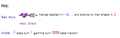
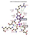
 LIGPLOT 2cfsA Mg1296.jpg 440 × 437; 26 KB
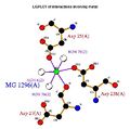
 LIGPLOT 2cfsA Mg1297.jpg 391 × 297; 17 KB
 LIGPLOT 2cftA.jpg 468 × 499; 35 KB
- LOC144557(with ligand bound).doc ; 128 KB
- LargeSimilarsequences3.txt ; 29 KB
- Lecture1.pdf 0 × 0; 1.64 MB
- Lecture2.pdf 0 × 0; 654 KB
- Lecture3.pdf 0 × 0; 1.68 MB
- Lecture4.pdf 0 × 0; 1,012 KB
- Lecture5.pdf 0 × 0; 181 KB
- Lecture6.pdf 0 × 0; 1.86 MB
- Lecture 1 2007 mac red.pdf 0 × 0; 4.02 MB
- Lee1971 JMolBiol55-379.pdf 0 × 0; 2.57 MB
- Lemkul ABB2007.pdf 0 × 0; 1,000 KB
 Ligand.JPG 759 × 610; 58 KB
 Ligand.jpg 451 × 306; 20 KB
 Ligand.png 744 × 785; 59 KB
 Ligand Qsite.png 640 × 480; 88 KB
 Ligand of bovine.png 400 × 400; 4 KB
 Ligand structure visualisation.jpg 1,161 × 827; 280 KB
 Ligand structure visualisation .jpg 1,161 × 827; 280 KB
 Ligand stucture and environment.png 1,347 × 416; 209 KB
 Ligandbindingsitepredictioni2.png 576 × 706; 31 KB
- Lindahl2000 JPhysChem113-3882.pdf 0 × 0; 565 KB
 Linker.png 640 × 434; 166 KB
- Liu MscL-2009.pdf 0 × 0; 1.15 MB
 Localisationhuman.jpg 520 × 790; 107 KB
 Localisationmouse.jpg 520 × 610; 87 KB
 MAS2.png 923 × 92; 10 KB
 MAS3.png 926 × 92; 11 KB
 MDgroup.JPG 480 × 360; 133 KB
 MDgroup.jpg 480 × 360; 133 KB
 MDgroup2.JPG 480 × 360; 144 KB
 MG1.jpg 800 × 300; 8 KB
 MG2-.jpg 800 × 300; 7 KB
 MG2.jpg 800 × 300; 7 KB
 MG 400.gif 400 × 400; 805 bytes
 MSA.JPG 1,280 × 1,024; 514 KB
 MSA.png 1,265 × 636; 120 KB
 MSA1.png 927 × 92; 12 KB
 MSA11.jpg 1,280 × 889; 116 KB
 MSA2.jpg 1,267 × 850; 193 KB
 MSA3.jpg 1,264 × 847; 161 KB
 MSA4.jpg 1,264 × 849; 131 KB
 MSA4.png 919 × 92; 11 KB
 MSA5.jpg 1,267 × 847; 199 KB
 MSA5.png 926 × 92; 12 KB
 MSA6.jpg 1,263 × 849; 151 KB
 MSA6.png 929 × 92; 10 KB
 MSA7.jpg 1,267 × 844; 128 KB
 MSA7.png 929 × 92; 10 KB
 MSA8.jpg 1,266 × 850; 101 KB
- MSAAnnotated.aln ; 59 KB
 MSAfigure2.JPG 899 × 864; 335 KB
 MSAfigure2.jpg 899 × 864; 335 KB
 MSE.png 309 × 165; 17 KB
 MSE 400.gif 400 × 400; 2 KB
- Mahoney2000 TIP5P.pdf 0 × 0; 184 KB
 Mark group 2.JPG 1,026 × 682; 362 KB
- Martin BMCStructBiol05.pdf 0 × 0; 960 KB
- Mayer2004 ChemPhysLett393-209.pdf 0 × 0; 170 KB
 Md todo.jpg 37 × 20; 1 KB
 Mdgroup2.jpg 480 × 360; 144 KB
 Mega.jpg 502 × 331; 34 KB
- Mel's picture of secondary str..bmp 0 × 0; 906 KB
 Mel's picture of secondary str..jpg 579 × 533; 69 KB
 Metallo.jpg 399 × 127; 20 KB
- Mixture models.pdf 0 × 0; 320 KB
- Modeling errors in NOE.pdf 0 × 0; 88 KB
- MolecularSurfaces.pdf 0 × 0; 6.39 MB
 Motif.jpg 754 × 472; 63 KB
 Motif 1 and 2.jpg 1,024 × 748; 199 KB
 Motif 3 and 4.jpg 1,024 × 748; 200 KB
 Motif Sequence1.JPG 1,149 × 525; 108 KB
 Motif Sequence2.JPG 657 × 218; 24 KB
 Motif alignment.jpg 724 × 100; 31 KB
 MouseSymatlas.png 520 × 610; 13 KB
 Mouse gene atlas.png 520 × 610; 13 KB
 Mousepseudomonasalign.jpg 564 × 827; 110 KB
 Movie.jpg 640 × 480; 73 KB
 Movie0001.png 640 × 480; 74 KB
- Msa1.bmp 0 × 0; 1.81 MB
 Msa1.jpg 1,141 × 554; 264 KB
 Msa16.png 1,013 × 500; 572 KB
 Msa17.png 998 × 490; 464 KB
 Msa2.jpg 1,141 × 559; 291 KB
 Msa3.jpg 1,141 × 554; 277 KB
 Msafinal.png 1,268 × 846; 716 KB
 Msaseg1a.JPG 863 × 433; 144 KB
 Msaseg1b.PNG 947 × 475; 148 KB
 Msaseg1b.png 947 × 475; 148 KB
 Msaseg2.png 948 × 474; 244 KB
 Msaseg3.png 947 × 484; 214 KB
 Msatree2.png 977 × 576; 87 KB
 Msatree2ece.png 932 × 565; 93 KB
 Msatree3.png 979 × 575; 90 KB
- MsbA Chang01.pdf 0 × 0; 1.66 MB
- MsbA Jeffrey09.pdf 0 × 0; 706 KB
- MsbA Miller06.pdf 0 × 0; 270 KB


