Summary Function for Report: Difference between revisions
No edit summary |
No edit summary |
||
| Line 1: | Line 1: | ||
<div class="Section1"> | |||
[[Image: | <span lang="EN-GB" style="mso-ansi-language: EN-GB"><font color="black"><font face=""Lucida Grande""></font></font></span> | ||
<center><span lang="EN-GB" style="mso-ansi-language: EN-GB"><font color="black"><font face=""Lucida Grande"">'''LOC55471 is a highly conserved non-nuclear Methyltransferase: '''</font></font></span></center> | |||
<center><span lang="EN-GB" style="mso-ansi-language: EN-GB"><font color="black"><font face=""Lucida Grande"">'''a functional analysis'''</font></font></span></center> | |||
<span lang="EN-GB" style="mso-ansi-language: EN-GB"><font color="black"><font face=""Lucida Grande""></font></font></span> | |||
<span lang="EN-GB" style="mso-ansi-language: EN-GB"><font color="black"><font face=""Lucida Grande"">By using ProFunc (Laskowski ''et al''</font></font></span><span lang="EN-GB" style="mso-ansi-language: EN-GB"><font color="black"><font face=""Lucida Grande"">, 2005) the most likely biochemical function of the unknown bacterial Protein 1zkd was determined as Methyltransferase. </font></font></span> | |||
<span lang="EN-GB" style="mso-ansi-language: EN-GB"><font color="black"><font face=""Lucida Grande""></font></font></span> | |||
<span lang="EN-GB" style="mso-ansi-language: EN-GB"><font color="black"><font face=""Lucida Grande"">Matching structures were determined by SSM Secondary Structure Matching (Krissinel & Henrick, 2004) showing possible matches with 9 Methyltransferases from both human and bacteria (Fig.1).</font></font></span> | |||
<span lang="EN-GB" style="mso-ansi-language: EN-GB"><font color="black"><font face=""Lucida Grande""></font></font></span> | |||
<center><span lang="EN-GB" style="mso-ansi-language: EN-GB"><font color="black"><font face=""Lucida Grande"">[[Image:image002.jpg]]</font></font></span></center> | |||
<span lang="EN-GB" style="mso-ansi-language: EN-GB"><font color="black"><font face=""Lucida Grande"">Figure 1. SSM results showing ten sequences with a sequences id around 20 % with higher matching folds. </font></font></span> | |||
<span lang="EN-GB" style="mso-ansi-language: EN-GB"><font color="black"><font face=""Lucida Grande""></font></font></span> | |||
<span lang="EN-GB" style="mso-ansi-language: EN-GB"><font color="black"><font face=""Lucida Grande""></font></font></span> | |||
<span lang="EN-GB" style="mso-ansi-language: EN-GB"><font color="black"><font face=""Lucida Grande""></font></font></span> | |||
<span lang="EN-GB" style="mso-ansi-language: EN-GB"><font color="black"><font face=""Lucida Grande"">Ligand Template Matches LIG (Laskowski ''et al''</font></font></span><span lang="EN-GB" style="mso-ansi-language: EN-GB"><font color="black"><font face=""Lucida Grande"">,</font></font></span><span lang="EN-GB" style="mso-ansi-language: EN-GB"><font color="black"><font face=""Lucida Grande"">2005) revealed a probable match with the Protein-l-isoaspartate o-methyltransferase 1dl5 (Fig.2).</font></font></span> | |||
<span lang="EN-GB" style="mso-ansi-language: EN-GB"><font color="black"><font face=""Lucida Grande""></font></font></span> | |||
<center><span lang="EN-GB" style="mso-ansi-language: EN-GB"><font color="black"><font face=""Lucida Grande"">[[Image:image004.jpg]]</font></font></span></center> | |||
<span lang="EN-GB" style="mso-ansi-language: EN-GB"><font color="black"><font face=""Lucida Grande"">Figure 2. LIG results support the hypothesis of 1zkd being a methyltransferase.</font></font></span> | |||
<span lang="EN-GB" style="mso-ansi-language: EN-GB"><font color="black"><font face=""Lucida Grande""></font></font></span> | |||
<span lang="EN-GB" style="mso-ansi-language: EN-GB"><font color="black"><font face=""Lucida Grande""></font></font></span> | |||
<span lang="EN-GB" style="mso-ansi-language: EN-GB"><font color="black"><font face=""Lucida Grande""></font></font></span> | |||
<span lang="EN-GB" style="mso-ansi-language: EN-GB"><font color="black"><font face=""Lucida Grande""></font></font></span> | |||
<span lang="EN-GB" style="mso-ansi-language: EN-GB"><font color="black"><font face=""Lucida Grande""></font></font></span> | |||
<span lang="EN-GB" style="mso-ansi-language: EN-GB"><font color="black"><font face=""Lucida Grande""></font></font></span> | |||
<span lang="EN-GB" style="mso-ansi-language: EN-GB"><font color="black"><font face=""Lucida Grande"">REV Reverse Template Matches (Laskowski ''et al''</font></font></span><span lang="EN-GB" style="mso-ansi-language: EN-GB"><font color="black"><font face=""Lucida Grande"">, 2005) also showed probable matches for several methyltransferases (Fig.3).</font></font></span> | |||
<center><span lang="EN-GB" style="mso-ansi-language: EN-GB"><font color="black"><font face=""Lucida Grande"">[[Image:image006.jpg]]</font></font></span></center> | |||
<span lang="EN-GB" style="mso-ansi-language: EN-GB"><font color="black"><font face=""Lucida Grande"">Figure 3. REV results showing five probable matches, which are all methyl or dimethyltransferases.</font></font></span> | |||
<span lang="EN-GB" style="mso-ansi-language: EN-GB"><font color="black"><font face=""Lucida Grande""></font></font></span> | |||
<span lang="EN-GB" style="mso-ansi-language: EN-GB"><font color="black"><font face=""Lucida Grande"">Superfamily program searches against a library of Hidden Markov Models HMMs (Gough ''et al''</font></font></span><span lang="EN-GB" style="mso-ansi-language: EN-GB"><font color="black"><font face=""Lucida Grande"">, 2001; Madera ''et al''</font></font></span><span lang="EN-GB" style="mso-ansi-language: EN-GB"><font color="black"><font face=""Lucida Grande"">, 2004) derived from SCOP families revealed similarities to the superfamily S-Adenosylmethionine-dependent Methyltransferases (E-value 6.69e-06).</font></font></span> | |||
<span lang="EN-GB" style="mso-ansi-language: EN-GB"><font color="black"><font face=""Lucida Grande""></font></font></span> | |||
<span lang="EN-GB" style="mso-ansi-language: EN-GB"><font color="black"><font face=""Lucida Grande"">No DNA binding motifs (helix-turn-helix) were found in the ProFunc search.</font></font></span> | |||
<span lang="EN-GB" style="mso-ansi-language: EN-GB"><font color="black"><font face=""Lucida Grande""></font></font></span> | |||
<span lang="EN-GB" style="mso-ansi-language: EN-GB"><font color="black"><font face=""Lucida Grande"">Genomic context of 1zkd in the genome of </font></font></span><font color="black"><font face=""Lucida Grande""><font size="13.0pt">''Rhodopseudomonas palustris''</font></font></font><span style="mso-ansi-language: EN-GB"><font color="black"><font face=""Lucida Grande""></font></font></span><span lang="EN-GB" style="mso-ansi-language: EN-GB"><font color="black"><font face=""Lucida Grande"">from the NCBI Entrez Gene database shows a genomic co-localisation with another transferase, an oxidase, a kinase and another hypothetical protein (Fig.4).</font></font></span> | |||
<span lang="EN-GB" style="mso-ansi-language: EN-GB"><font color="black"><font face=""Lucida Grande""></font></font></span> | |||
<span lang="EN-GB" style="mso-ansi-language: EN-GB"><font color="black"><font face=""Lucida Grande"">[[Image:image008.jpg]]Figure 4. The RPA4359 gene of the protein 1zkd is co-located with an upstream prolipoprotein diacylglyceryl transferase gene (1gt) and downstream with a multicopper polyphenol oxidase (RPA4360), a ribose-phosphate pyrophosphokinase (ribP) and another hypothetical protein of unknown function gene (RPA4361).</font></font></span> | |||
<span lang="EN-GB" style="mso-ansi-language: EN-GB"><font color="black"><font face=""Lucida Grande""></font></font></span> | |||
<span lang="EN-GB" style="mso-ansi-language: EN-GB"><font color="black"><font face=""Lucida Grande""></font></font></span> | |||
<span lang="EN-GB" style="mso-ansi-language: EN-GB"><font color="black"><font face=""Lucida Grande""></font></font></span> | |||
<span lang="EN-GB" style="mso-ansi-language: EN-GB"><font color="black"><font face=""Lucida Grande"">Nucleo (Nuclear Protein Localisation Prediction) predicted a chance of 0.07 for the mouse ortholog and a chance of 0.20 for the human ortholog of 1zkd to be located in the nucleus (Hawkins ''et al''</font></font></span><span lang="EN-GB" style="mso-ansi-language: EN-GB"><font color="black"><font face=""Lucida Grande"">, 2006).</font></font></span> | |||
<span lang="EN-GB" style="mso-ansi-language: EN-GB"><font color="black"><font face=""Lucida Grande""></font></font></span> | |||
<span lang="EN-GB" style="mso-ansi-language: EN-GB"><font color="black"><font face=""Lucida Grande"">LOCATE data was available for the mouse ortholog showing that it is a soluble, non-secreted protein with higher scores for a localisation in mitochondria or the cytoplasm (Fink ''et al''</font></font></span><span lang="EN-GB" style="mso-ansi-language: EN-GB"><font color="black"><font face=""Lucida Grande"">, 2006).</font></font></span> | |||
<span lang="EN-GB" style="mso-ansi-language: EN-GB"><font color="black"><font face=""Lucida Grande""></font></font></span> | |||
<span lang="EN-GB" style="mso-ansi-language: EN-GB"><font color="black"><font face=""Lucida Grande"">Expression profile data of the mouse and human ortholog were suggested by analysis of EST counts from NCBI UniGene database (http://www.ncbi.nlm.nih.gov/sites/entrez?db=unigene). ESTs were found in diverse tissues including brain, liver, lung, muscle and endocrine system showing that the target protein is expressed in a wide range of different cells (Fig.5).</font></font></span> | |||
<span lang="EN-GB" style="mso-ansi-language: EN-GB"><font color="black"><font face=""Lucida Grande""></font></font></span> | |||
<span lang="EN-GB" style="mso-ansi-language: EN-GB"><font color="black"><font face=""Lucida Grande""></font></font></span> | |||
<span lang="EN-GB" style="mso-ansi-language: EN-GB"><font color="black"><font face=""Lucida Grande""></font></font></span> | |||
<span lang="EN-GB" style="mso-ansi-language: EN-GB"><font color="black"><font face=""Lucida Grande""></font></font></span> | |||
<center><span lang="EN-GB" style="mso-ansi-language: EN-GB"><font color="black"><font face=""Lucida Grande"">''''''</font></font></span></center> | |||
<center><span lang="EN-GB" style="mso-ansi-language: EN-GB"><font color="black"><font face=""Lucida Grande"">''''''</font></font></span></center> | |||
<center><span lang="EN-GB" style="mso-ansi-language: EN-GB"><font color="black"><font face=""Lucida Grande"">'''[[Image:image010.jpg]][[Image:image012.jpg]]'''</font></font></span></center> | |||
<span lang="EN-GB" style="mso-ansi-language: EN-GB"><font color="black"><font face=""Lucida Grande"">Figure 5. Expression profiles for the mouse and human ortholog of 1zkd suggested by analysis of EST counts.</font></font></span> | |||
<center><span lang="EN-GB" style="mso-ansi-language: EN-GB"><font color="black"><font face=""Lucida Grande"">''''''</font></font></span></center> | |||
<center><span lang="EN-GB" style="mso-ansi-language: EN-GB"><font color="black"><font face=""Lucida Grande"">''''''</font></font></span></center> | |||
<span lang="EN-GB" style="mso-ansi-language: EN-GB"><font color="black"><font face=""Lucida Grande""></font></font></span> | |||
<span lang="EN-GB" style="mso-ansi-language: EN-GB"><font color="black"><font face=""Lucida Grande""></font></font></span> | |||
<span lang="EN-GB" style="mso-ansi-language: EN-GB"><font color="black"><font face=""Lucida Grande""></font></font></span> | |||
<span lang="EN-GB" style="mso-ansi-language: EN-GB"><font color="black"><font face=""Lucida Grande"">Electrostatic properties and surface charges of 1zkd were modelled using Adaptive Poisson-Boltzmann Solver APBS (Baker ''et al''</font></font></span><span lang="EN-GB" style="mso-ansi-language: EN-GB"><font color="black"><font face=""Lucida Grande"">, 2001) and visualisation was performed by using Pymol (http://www.pymol.org). According to the resulting model, the 1zkd protein got a mostly negatively charged surface (Fig.6), indicating that interactions with the negatively charged backbone of nucleic acids are rather unlikely.</font></font></span> | |||
<span lang="EN-GB" style="mso-ansi-language: EN-GB"><font color="black"><font face=""Lucida Grande""></font></font></span> | |||
<span lang="EN-GB" style="mso-ansi-language: EN-GB"><font color="black"><font face=""Lucida Grande""></font></font></span> | |||
<span lang="EN-GB" style="mso-ansi-language: EN-GB"><font color="black"><font face=""Lucida Grande"">''''''</font></font></span> | |||
<center><span lang="EN-GB" style="mso-ansi-language: EN-GB"><font color="black"><font face=""Lucida Grande"">'''[[Image:image014.png]][[Image:image016.png]]'''</font></font></span></center> | |||
<center><span lang="EN-GB" style="mso-ansi-language: EN-GB"><font color="black"><font face=""Lucida Grande"">'''[[Image:image018.png]][[Image:image020.png]]'''</font></font></span></center> | |||
<span lang="EN-GB" style="mso-ansi-language: EN-GB"><font color="black"><font face=""Lucida Grande"">Figure 6: Surface charges of 1zkd as a dimer. Red colour indicating negative charges, blue colour indicating positive charges.</font></font></span> | |||
<span lang="EN-GB" style="mso-ansi-language: EN-GB"><font color="black"><font face=""Lucida Grande""></font></font></span> | |||
<span lang="EN-GB" style="mso-ansi-language: EN-GB"><font color="black"><font face=""Lucida Grande"">Taken all results together it can be assumed that the bacterial 1zkd and its mouse and human orthologs might act as methyltransferases in a variety of tissues. The substrate might be s-adenosylmethionine leaving s-adenosylhomocysteine as a product after transferring the reactive methyl group to a protein or nucleic acid (Fig.7).</font></font></span> | |||
<span lang="EN-GB" style="mso-ansi-language: EN-GB"><font color="black"><font face=""Lucida Grande"">The lack of nucleus localisation signals (NLS) in the orthologs and the negatively charged surface of 1zkd indicate that this conserved protein might rather act as a modifier of other proteins in the cytoplasm or mitochondria by transferring methyl groups from the substrate S-adenosylmethionine to other proteins. This posttranslational modification could have a variety of impacts on target protein function e.g. in cell signalling. Although it remains unclear how these protein-protein interaction might occur. </font></font></span> | |||
<span lang="EN-GB" style="mso-ansi-language: EN-GB"><font color="black"><font face=""Lucida Grande"">Another possibility could be that 1izkd and its orthologs are associated with other proteins while performing transmethylation, which might be able to bind nucleic acids like RNA. Thus, it cannot be excluded that the 1zkd and its orthologs might be involved in RNA methylation in the cytoplasm or in mitochondria (Fig.7).</font></font></span> | |||
<span lang="EN-GB" style="mso-ansi-language: EN-GB"><font color="black"><font face=""Lucida Grande"">''''''</font></font></span> | |||
<span lang="EN-GB" style="mso-ansi-language: EN-GB"><font color="black"><font face=""Lucida Grande"">''''''</font></font></span> | |||
<span lang="EN-GB" style="mso-ansi-language: EN-GB"><font color="black"><font face=""Lucida Grande"">''''''</font></font></span> | |||
<center><span lang="EN-GB" style="mso-ansi-language: EN-GB"><font color="black"><font face=""Lucida Grande"">'''[[Image:image022.jpg]]'''</font></font></span></center> | |||
<span lang="EN-GB" style="mso-ansi-language: EN-GB"><font color="black">Figure 7. Possible functions of 1zkd and its orthologs in the cytoplasm or mitochondrium. They might transfer reactive methyl-groups to other proteins (X) or might build complexes with other protein (Y) to methylate RNA.</font></span> | |||
<span lang="EN-GB" style="mso-ansi-language: EN-GB"><font color="black">''''''</font></span> | |||
<span style="mso-ignore: vglayout; position: absolute; z-index: 2; left: 0px; margin-left: 21px; margin-top: 9px; width: 3px; height: 3px">[[Image:image023.png]]</span><span style="mso-ignore: vglayout; position: absolute; z-index: 1; left: 0px; margin-left: 21px; margin-top: 9px; width: 3px; height: 3px">[[Image:image024.png]]</span><span lang="EN-GB" style="mso-ansi-language: EN-GB"><font color="black">''''''</font></span> | |||
{| align="left" | |||
|- | |||
| style="vertical-align: top; background: white" width="56" height="29" bgcolor="white" align="left" valign="top" | <span style="position: absolute; z-index: 1">{| width="100%" | |||
| | |||
<div class="shape" style="padding: 3.6pt 7.2pt 3.6pt 7.2pt"><font size="10.0pt"></font></div> | |||
|}</span> | |||
|} | |||
<span lang="EN-GB" style="mso-ansi-language: EN-GB"><font color="black"><font face=""Lucida Grande"">''''''</font></font></span> | |||
<center><span lang="EN-GB" style="mso-ansi-language: EN-GB"><font color="black"><font face=""Lucida Grande"">''''''</font></font></span></center> | |||
<br style="mso-ignore: vglayout" clear="all" /> | |||
<center><span lang="EN-GB" style="mso-ansi-language: EN-GB"><font color="black"><font face=""Lucida Grande"">'''References'''</font></font></span></center> | |||
<span lang="EN-GB" style="mso-ansi-language: EN-GB"><font color="black"><font face=""Lucida Grande""></font></font></span> | |||
<font color="black"><font face=""Lucida Grande""><font size="13.0pt">Baker, N.A., Sept, D., Joseph, S., Holst, M.J. & McCammon, J.A. (2001). Electrostatics of nanosystems: application to microtubules and the ribosome. ''Proc. Natl. Acad. Sci., ''</font></font></font><font color="black"><font face=""Lucida Grande""><font size="13.0pt">'''98'''</font></font></font><font color="black"><font face=""Lucida Grande""><font size="13.0pt">, 10037-10041.</font></font></font> | |||
<span lang="EN-GB" style="mso-ansi-language: EN-GB"><font color="black"><font face=""Lucida Grande""></font></font></span> | |||
<font color="black"><font face=""Lucida Grande""><font size="13.0pt">DeLano, W.L. (2002). The PyMOL Molecular Graphics System. DeLano Scientific, Palo Alto, CA, USA.</font></font></font> | |||
<span lang="EN-GB" style="mso-ansi-language: EN-GB"><font color="black"><font face=""Lucida Grande""></font></font></span> | |||
<font color="black"><font face=""Lucida Grande"">Fink, J.L., Aturaliya, R.N., Davis, M.J., Zhang, F., Hanson, K., Teasdale, M.S., Kai, C., Kawai, J., Carninci, P., Hayashizaki, Y. & Teasdale, R.D. (2006). LOCATE: a mouse protein subcellular localization database. ''Nucleic Acids Res.''</font></font><font color="black"><font face=""Lucida Grande"">, '''34'''</font></font><font color="black"><font face=""Lucida Grande"">(Database issue),D213-7</font></font><font color="black"><font face=""Lucida Grande""><font size="13.0pt">.</font></font></font> | |||
<font color="black"><font face=""Lucida Grande""><font size="13.0pt"></font></font></font> | |||
<font color="black"><font face=""Lucida Grande""><font size="13.0pt">Gough, J., Karplus, K., Hughey, R. & Chothia, C. (2001). Assignment of homology to genome sequences using a library of Hidden Markov Models that represent all proteins of known structure. ''J. Mol. Biol''</font></font></font><font color="black"><font face=""Lucida Grande""><font size="13.0pt">., '''313'''</font></font></font><font color="black"><font face=""Lucida Grande""><font size="13.0pt">, 903-919.</font></font></font> | |||
<font color="black"><font face=""Lucida Grande""><font size="13.0pt"></font></font></font> | |||
<font color="black"><font face=""Lucida Grande""><font size="13.0pt">Hawkins, J., Davis, L. & BodŽn, M. (2006). Predicting Nuclear Proteins. Manuscript Submitted to ''Bioinformatics''</font></font></font><font color="black"><font face=""Lucida Grande""><font size="13.0pt">.</font></font></font> | |||
<font color="black"><font face=""Lucida Grande""><font size="13.0pt"></font></font></font> | |||
<font color="black"><font face=""Lucida Grande""><font size="13.0pt">Krissinel, E. & Henrick, K. (2004). Secondary-structure matching (SSM), a new tool for fast protein structure alignment in three dimensions. ''Acta Cryst.''</font></font></font><font color="black"><font face=""Lucida Grande""><font size="13.0pt">, '''D60'''</font></font></font><font color="black"><font face=""Lucida Grande""><font size="13.0pt">, 2256-2268.</font></font></font> | |||
<font color="black"><font face=""Lucida Grande""><font size="13.0pt"></font></font></font> | |||
<font color="black"><font face=""Lucida Grande""><font size="13.0pt">Laskowski, R. A., Watson, J. D. & Thornton, J. M. (2005). ProFunc: a server for predicting protein function from 3D structure. ''Nucleic Acids Res.''</font></font></font><font color="black"><font face=""Lucida Grande""><font size="13.0pt">, '''33'''</font></font></font><font color="black"><font face=""Lucida Grande""><font size="13.0pt">, W89-W93.</font></font></font> | |||
<font color="black"><font face=""Lucida Grande""><font size="13.0pt"></font></font></font> | |||
<font color="black"><font face=""Lucida Grande""><font size="13.0pt">Laskowski, R.A., Watson, J.D. & Thornton, J.M. (2005). Protein function prediction using local 3D templates. ''J. Mol. Biol''</font></font></font><font color="black"><font face=""Lucida Grande""><font size="13.0pt">., '''351'''</font></font></font><font color="black"><font face=""Lucida Grande""><font size="13.0pt">, 614-626.</font></font></font> | |||
<font color="black"><font face=""Lucida Grande""><font size="13.0pt"></font></font></font> | |||
<font color="black"><font face=""Lucida Grande""><font size="13.0pt">Madera, M., Vogel, C., Kummerfeld, S.K., Chothia, C. & Gough, J. (2004). The SUPERFAMILY database in 2004: additions and improvements. ''Nucl. Acids Res''</font></font></font><font color="black"><font face=""Lucida Grande""><font size="13.0pt">., '''32'''</font></font></font><font color="black"><font face=""Lucida Grande""><font size="13.0pt">, D235-D239.</font></font></font> | |||
</div> | |||
Revision as of 04:47, 9 June 2007
By using ProFunc (Laskowski et al, 2005) the most likely biochemical function of the unknown bacterial Protein 1zkd was determined as Methyltransferase.
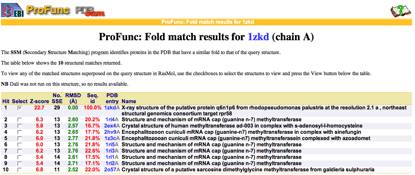
Matching structures were determined by SSM Secondary Structure Matching (Krissinel & Henrick, 2004) showing possible matches with 9 Methyltransferases from both human and bacteria (Fig.1).

Figure 1. SSM results showing ten sequences with a sequences id around 20 % with higher matching folds.
Ligand Template Matches LIG (Laskowski et al,2005) revealed a probable match with the Protein-l-isoaspartate o-methyltransferase 1dl5 (Fig.2).
Figure 2. LIG results support the hypothesis of 1zkd being a methyltransferase.
REV Reverse Template Matches (Laskowski et al, 2005) also showed probable matches for several methyltransferases (Fig.3).
Figure 3. REV results showing five probable matches, which are all methyl or dimethyltransferases.
Superfamily program searches against a library of Hidden Markov Models HMMs (Gough et al, 2001; Madera et al, 2004) derived from SCOP families revealed similarities to the superfamily S-Adenosylmethionine-dependent Methyltransferases (E-value 6.69e-06).
No DNA binding motifs (helix-turn-helix) were found in the ProFunc search.
Genomic context of 1zkd in the genome of Rhodopseudomonas palustrisfrom the NCBI Entrez Gene database shows a genomic co-localisation with another transferase, an oxidase, a kinase and another hypothetical protein (Fig.4).
File:Image008.jpgFigure 4. The RPA4359 gene of the protein 1zkd is co-located with an upstream prolipoprotein diacylglyceryl transferase gene (1gt) and downstream with a multicopper polyphenol oxidase (RPA4360), a ribose-phosphate pyrophosphokinase (ribP) and another hypothetical protein of unknown function gene (RPA4361).
Nucleo (Nuclear Protein Localisation Prediction) predicted a chance of 0.07 for the mouse ortholog and a chance of 0.20 for the human ortholog of 1zkd to be located in the nucleus (Hawkins et al, 2006).
LOCATE data was available for the mouse ortholog showing that it is a soluble, non-secreted protein with higher scores for a localisation in mitochondria or the cytoplasm (Fink et al, 2006).
Expression profile data of the mouse and human ortholog were suggested by analysis of EST counts from NCBI UniGene database (http://www.ncbi.nlm.nih.gov/sites/entrez?db=unigene). ESTs were found in diverse tissues including brain, liver, lung, muscle and endocrine system showing that the target protein is expressed in a wide range of different cells (Fig.5).
Figure 5. Expression profiles for the mouse and human ortholog of 1zkd suggested by analysis of EST counts.
Electrostatic properties and surface charges of 1zkd were modelled using Adaptive Poisson-Boltzmann Solver APBS (Baker et al, 2001) and visualisation was performed by using Pymol (http://www.pymol.org). According to the resulting model, the 1zkd protein got a mostly negatively charged surface (Fig.6), indicating that interactions with the negatively charged backbone of nucleic acids are rather unlikely.
'
Figure 6: Surface charges of 1zkd as a dimer. Red colour indicating negative charges, blue colour indicating positive charges.
Taken all results together it can be assumed that the bacterial 1zkd and its mouse and human orthologs might act as methyltransferases in a variety of tissues. The substrate might be s-adenosylmethionine leaving s-adenosylhomocysteine as a product after transferring the reactive methyl group to a protein or nucleic acid (Fig.7).
The lack of nucleus localisation signals (NLS) in the orthologs and the negatively charged surface of 1zkd indicate that this conserved protein might rather act as a modifier of other proteins in the cytoplasm or mitochondria by transferring methyl groups from the substrate S-adenosylmethionine to other proteins. This posttranslational modification could have a variety of impacts on target protein function e.g. in cell signalling. Although it remains unclear how these protein-protein interaction might occur.
Another possibility could be that 1izkd and its orthologs are associated with other proteins while performing transmethylation, which might be able to bind nucleic acids like RNA. Thus, it cannot be excluded that the 1zkd and its orthologs might be involved in RNA methylation in the cytoplasm or in mitochondria (Fig.7).
'
'
'

Figure 7. Possible functions of 1zkd and its orthologs in the cytoplasm or mitochondrium. They might transfer reactive methyl-groups to other proteins (X) or might build complexes with other protein (Y) to methylate RNA.
'
File:Image023.pngFile:Image024.png'
| {| width="100%" |
|}
'
Baker, N.A., Sept, D., Joseph, S., Holst, M.J. & McCammon, J.A. (2001). Electrostatics of nanosystems: application to microtubules and the ribosome. Proc. Natl. Acad. Sci., 98, 10037-10041.
DeLano, W.L. (2002). The PyMOL Molecular Graphics System. DeLano Scientific, Palo Alto, CA, USA.
Fink, J.L., Aturaliya, R.N., Davis, M.J., Zhang, F., Hanson, K., Teasdale, M.S., Kai, C., Kawai, J., Carninci, P., Hayashizaki, Y. & Teasdale, R.D. (2006). LOCATE: a mouse protein subcellular localization database. Nucleic Acids Res., 34(Database issue),D213-7.
Gough, J., Karplus, K., Hughey, R. & Chothia, C. (2001). Assignment of homology to genome sequences using a library of Hidden Markov Models that represent all proteins of known structure. J. Mol. Biol., 313, 903-919.
Hawkins, J., Davis, L. & BodŽn, M. (2006). Predicting Nuclear Proteins. Manuscript Submitted to Bioinformatics.
Krissinel, E. & Henrick, K. (2004). Secondary-structure matching (SSM), a new tool for fast protein structure alignment in three dimensions. Acta Cryst., D60, 2256-2268.
Laskowski, R. A., Watson, J. D. & Thornton, J. M. (2005). ProFunc: a server for predicting protein function from 3D structure. Nucleic Acids Res., 33, W89-W93.
Laskowski, R.A., Watson, J.D. & Thornton, J.M. (2005). Protein function prediction using local 3D templates. J. Mol. Biol., 351, 614-626.
Madera, M., Vogel, C., Kummerfeld, S.K., Chothia, C. & Gough, J. (2004). The SUPERFAMILY database in 2004: additions and improvements. Nucl. Acids Res., 32, D235-D239.