|
|
| Line 1: |
Line 1: |
| General structure of Pyridoxal Phosphatase<BR>
| | [[Image:2cftA Structure breakdown.jpg|left|thumb|500px|Fig 1. Courtesy (and edited) of Almo et al., 2007. Structure of 2cftA - Human Pyridoxal 5'-Phosphate Phosphatase with its substrate. As mentioned in the "Introduction" section, the core domain consists of beta sheets sandwiched by alpha helices. 2cftA is similar to 2cfsA, with the differences being the presence of a PLP ligand and the type of metal ions (2cfsA has magnesium ions, 2cftA has calcium ions). Notice the similarities between the structure of 2cftA and the PyMOL-generated image of 2cfsA (Fig 2.).]] |
| [[Image:2cftA Structure breakdown.jpg|left|thumb|500px|Courtesy (and edited) of Almo et al., 2007. This structure is actually that of 2cftA - Human Pyridoxal 5'-Phosphate Phosphatase with its substrate. It is similar to 2cfsA, with the differences being the presence of a PLP ligand and the type of metal ions (2cfsA has magnesium ions, 2cftA has calcium ions). Notice the similarities between the structure of 2cftA and 2cfsA]][[Image:2cfsA_PyMOL.png|left|thumb|500px|The three-dimensional structure of 2cfsA, as generated by PyMOL<BR> | | [[Image:2cfsA_PyMOL.png|left|thumb|500px|Fig 2. The three-dimensional structure of 2cfsA, as generated by PyMOL<BR> |
| <BR> | | <BR> |
| Red - Helix<BR> | | Red - Helix<BR> |
| Yellow - Sheet<BR> | | Yellow - Sheet<BR> |
| Green - Loop <BR>]] | | Green - Loop <BR> |
| | | Green Dots - Water molecules]] |
| <BR><BR><BR><BR><BR><BR><BR><BR><BR><BR><BR><BR><BR><BR><BR><BR><BR><BR><BR><BR><BR><BR><BR><BR><BR><BR><BR> | | <BR><BR><BR><BR><BR><BR><BR><BR><BR><BR><BR><BR><BR><BR><BR><BR><BR><BR><BR><BR><BR><BR><BR><BR><BR><BR><BR><BR> |
|
| |
|
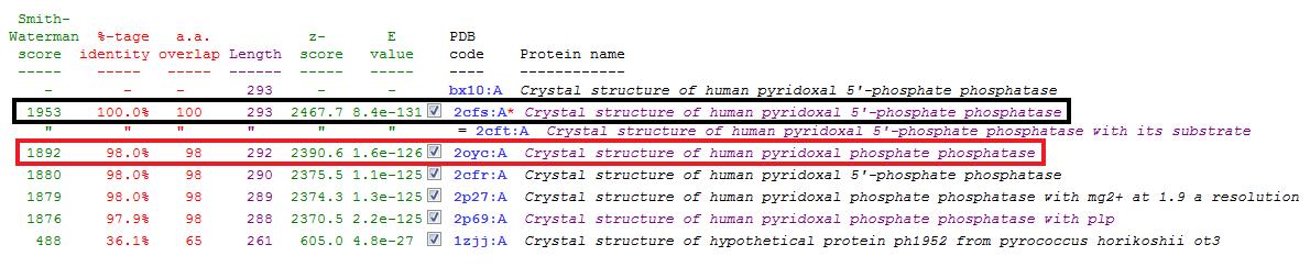
| '''DALI - 1st Run''' | | '''DALI - 1st Run''' |
Revision as of 15:19, 7 June 2008

Fig 1. Courtesy (and edited) of Almo et al., 2007. Structure of 2cftA - Human Pyridoxal 5'-Phosphate Phosphatase with its substrate. As mentioned in the "Introduction" section, the core domain consists of beta sheets sandwiched by alpha helices. 2cftA is similar to 2cfsA, with the differences being the presence of a PLP ligand and the type of metal ions (2cfsA has magnesium ions, 2cftA has calcium ions). Notice the similarities between the structure of 2cftA and the PyMOL-generated image of 2cfsA (Fig 2.).
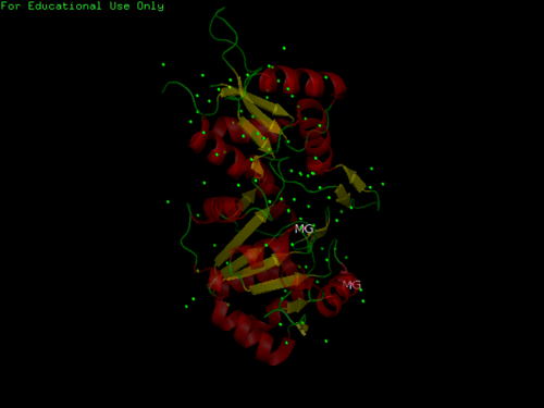
Fig 2. The three-dimensional structure of 2cfsA, as generated by PyMOL
Red - Helix
Yellow - Sheet
Green - Loop
Green Dots - Water molecules
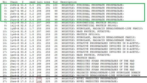
DALI - 1st Run
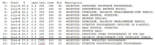
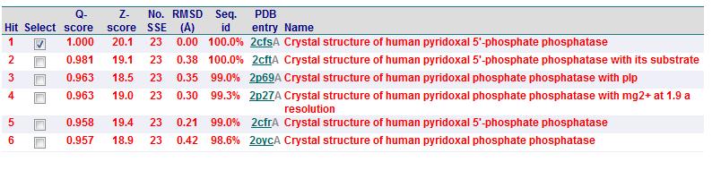
Significant hits as generated from the DALI database
Based on the results of the first run, how similar is 2oycA to 2cfsA? With the aid of PyMOL, 2oycA was super-imposed against 2cfsA...
Comparing 2oycA against 2cfsA
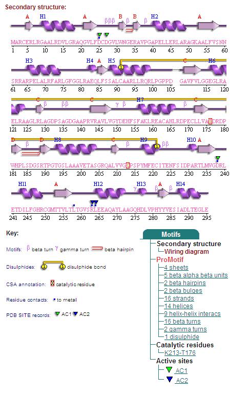
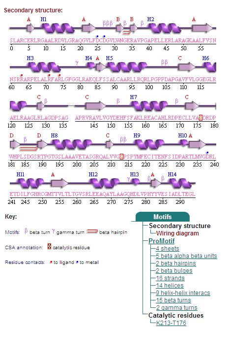
1. Secondary Structure
2. Topology Diagram
Topology Diagram of 2cfsA
Topology diagram of 2oycA
PROFUNC
Related Protein Sequences in the PDB (SAS)
Matches to existing PDB Structures
Secondary Structure Matching (SSM)
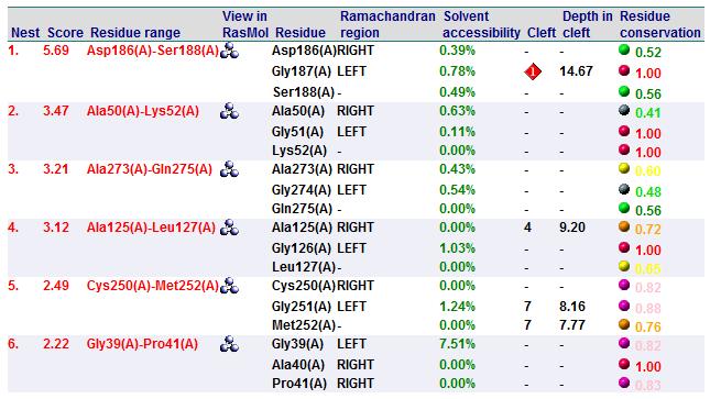
Nest Analysis
Summary of Predicted Function

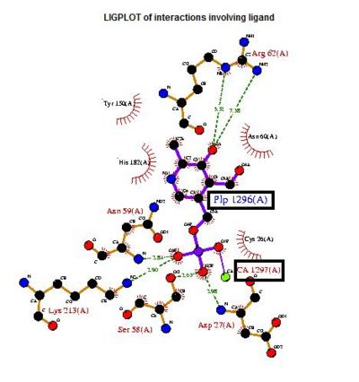
The LIGPLOT of interactions involving the PLP ligand in 2cftA. This was hypothesized to be the location of 2cftA's active site. The Mg 1296(A) ion of 2cfsA is located in the same position as the calcium ion of 2cftA. Therefore, there could be a possibility that the active site of 2cfsA could be at Mg 1296(A).
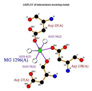
The LIGPLOT of interactions involving the Mg 1296(A) ion in 2cfsA. Notice the similarity of its location with relation to the calcium ion in 2cftA
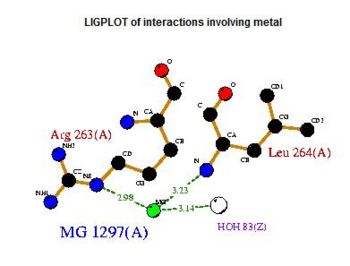
The LIGPLOT of interactions involving the Mg 1297(A) ion in 2cfsA. While it does not seem to share any similarities with the PLP ligand in 2cftA, the information provided by the LIGPLOT will be useful when visualizing the catalytic site of 2cfsA via PyMOL.
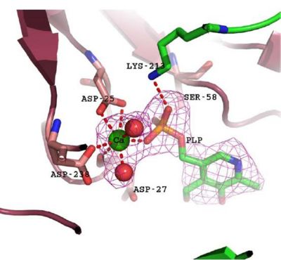
The catalytic site of 2cftA, with its PLP ligand and inhibitory calcium ion. As illustrated, the calcium ion is hepta-coordinated and participates in a bidentate interaction with the active site nucleophile Asp-25. Diagram and caption courtesy of Almo et al., 2007.
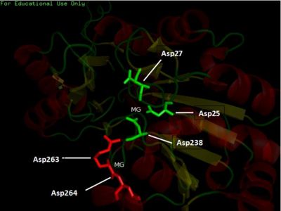
The three-dimensional visualization of the catalytic site of 2cfsA. This was generated based on the location of the respective Asp, as provided in Figs 10 and 11.
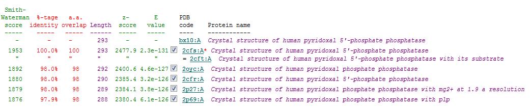
Dali - 2nd Run
The results obtained when 2cfsA was re-run against the DALI database
The second DALI run does not affect the results as PROFUNC has already indicated 2cftA to be the most closely-related to 2cfsA. On the contrary, the results serve as a confirmation that 2cfsA is indeed a Pyridoxal Phosphatase and belongs to the same family as 2oycA and 2cftA, 2 proteins that have been studied to aid in the structural and functional prediction of 2cfsA.
Back To Main Page


