Structure Protein: Difference between revisions
No edit summary |
|||
| Line 6: | Line 6: | ||
(chain A= green, chain B= cyan, chain c= purple, and chain d= yellow) | (chain A= green, chain B= cyan, chain c= purple, and chain d= yellow) | ||
Two molecular components were observed: | |||
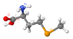
sulfate ion (SO4)[[Image:Sulfate ion.png]] and selenomethionine (MSE)[[Image:MSE.png]] | |||
Ten sulfate ions were observed. Each ion differs in arrangement as well as orientation related with each chains. | Ten sulfate ions were observed. Each ion differs in arrangement as well as orientation related with each chains. | ||
Revision as of 06:24, 5 June 2007
2ifu has 4 chains (A, B, C and D) integrated together to form the whole molecular structure as shown below:

(chain A= green, chain B= cyan, chain c= purple, and chain d= yellow)
Two molecular components were observed:
sulfate ion (SO4)and selenomethionine (MSE)


Ten sulfate ions were observed. Each ion differs in arrangement as well as orientation related with each chains.
protein sequence (FASTA)
>gi|118138356|pdb|2IFU|A Chain A, Crystal Structure Of A Gamma-Snap From Danio Rerio AIAAQKISEAHEHIAKAEKYLKTSFXKWKPDYDSAASEYAKAAVAFKNAKQLEQAKDAYLQEAEAHANNR SLFHAAKAFEQAGXXLKDLQRXPEAVQYIEKASVXYVENGTPDTAAXALDRAGKLXEPLDLSKAVHLYQQ AAAVFENEERLRQAAELIGKASRLLVRQQKFDEAAASLQKEKSXYKEXENYPTCYKKCIAQVLVQLHRAD YVAAQKCVRESYSIPGFSGSEDCAALEDLLQAYDEQDEEQLLRVCRSPLVTYXDNDYAKLAISLKVPGGG GGKKKPSASASAQPQEEEDDEYAGGLC
Strutural comparisons
Based on Dali search, 6 proteins with highest Z-value were chosen: 1qqe-A (protein transport), 2fi7-A (protein binding), 1a17 (hydrolase), 2ak6-A, 1fch-A (signalling protein), 1haz4-A (transcription factor),
snap-gamma: multiple structure alignment
Snap-gamma: sequence alignment with 1QQE:A

Dark colors: 2ifu:A
Light colors: 1qqe:A
SCOP
multiple alignment results, click here

Superimposed structure between 2ifuA; 2ifuB; 2ifuC; 2ifuD; 1qqeA; and 1b89A
Protein families
has 2 domains: Pfam at Sanger
alignment with:
Pfam-B_7270 (7-66)
highly conserved residue:
Pfam-B_15198 (87-307)
highly conserved residue :residue 104, 109, 121, 168
scorecons result indicated a Diversity of position scores: 89.4% where 100%=high diversity
InterProScan Results

links
CRYSTAL STRUCTURE OF THE VESICULAR TRANSPORT PROTEIN SEC17 1QQE
CLATHRIN HEAVY CHAIN PROXIMAL LEG SEGMENT (BOVINE) 1B89
http://myhits.isb-sib.ch/cgi-bin/motif_scan
http://www.ncbi.nlm.nih.gov/Structure/vast/vastsrv.cgi?sdid=181604