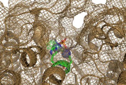
View of the Binding Pocket: Difference between revisions
| Line 5: | Line 5: | ||
==Ligand Interactions== | ==Ligand Interactions== | ||
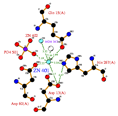
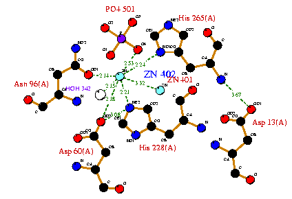
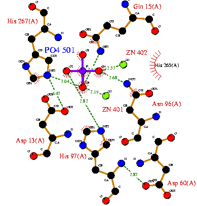
Below are the interactions between the two Zn atoms and the PO<sub>4</sub> ion in the binding pocket. | Below are the interactions between the two Zn atoms and the PO<sub>4</sub> ion in the binding pocket. Some of the residues shown here are conserved or invariant, such as Asp96 and His265. | ||
[[Image:Zn1 interactions.png]] | [[Image:Zn1 interactions.png]] | ||
[[Image:Zn2 interactions.png]] | [[Image:Zn2 interactions.png]] | ||
[[Image:PO4 interactions.png]] | [[Image:PO4 interactions.png]] | ||
Revision as of 01:36, 24 May 2008
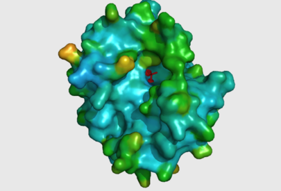
The large cavity of the 2nxf protein shown in a surface representation (left), showing its relative position with the rest of the protein. The ligands are shown here in red. The ribbon view, with mesh surface superimposed on top (right) is a close up of this same binding pocket, viewed from the right side of the protein. Analysis of the Dali results showed that the top 5 sequences contained three unchanging amino acid residues at positions 95 to 97 with a highly conserved acidic amino acid at position 98. The side-chains of conserved residues are shown here as coloured sticks. The two Zn atoms (spheres) appear to be indented into the surface of the protein with the phosphorous ion ligand sitting just above them on the surface of the protein.
Ligand Interactions
Below are the interactions between the two Zn atoms and the PO4 ion in the binding pocket. Some of the residues shown here are conserved or invariant, such as Asp96 and His265.
