DnaB Helicase
Introduction to Helicases
DNA usually exists as a double stranded duplex, but during DNA replication the strands of the double helix must be unwound and separated to form single-stranded DNA intermediates. Separation is carried out by molecular motors known as DNA helicases that move along the length of the DNA lattice. They move along the length of the DNA, destabilizing the interactions between complementary base pairs. The movement along the lattice and the separation of the DNA strands are coupled to the hydrolysis of nucleoside-5'-triphosphates. Helicases have the ability to move along the DNA lattice for long distances without dissociating. This is termed processive movement, and helicases have a high processibility. Processive movement is essential for helicases involved in DNA replication.
Helicases have at least 2 structural and functional strategies for achieving high processibility. Certain hexameric helicases form ring like structures that completely encircles at least one of the strands of a DNA duplex. Other helicases, Rep helicase from E. coli, are homodimeric and move processively along the DNA helix by means of a "hand-over-hand" movement. A key feature of "hand-over-hand" movement of a dimeric motor protein along a polymer is that at least one of the motor subunits must be bound to the polymer at any moment.
DnaB helicase is the major replicative DNA helicase in E. coli. It is a member of the hexameric DNA helicase family whose members are involved in DNA replication such as T4 DNA helicase, T7 DNA helicase, and SV40 T-antigen. In addition to their hexameric structure, these helicases show a common polarity of movement, and all except T-antigen have a 5' -> 3' polarity. DnaB helicase is a true multifunctional enzyme with a number of distinct enzymatic activities including ATP1 hydrolysis, DNA binding, and association with other replication proteins, such as DnaG primase and DNA polymerase III holoenzyme. The interactions with replication proteins in the replication fork allow it to form the replisome with primase and holoenzyme dimer in order to replicate the leading and lagging strands simultaneously. The energy of DNA-dependent ATP hydrolysis allows duplex DNA unwinding by the DnaB helicase and movement of the replisome synchronously with the progression of DNA unwinding and fork movement. DnaB helicase forms a hexamer which is partially Mg2+ dependent. DnaB helicase consists of 471 amino acid residues.
Function of DnaB Helicase
The function of DnaB helicase is to couple ATP hydrolysis with the unwinding of duplex DNA at the replication fork. This creates 2 anti parallel DNA single strands. The leading ssDNA polymer is the template for DNA polymerase III holoenzyme which synthesizes a continuous strand. The tao subunit of DNA polymerase III works by stabilizing the hexameric structure of DnaB helicase. The tao subunit must bind to DNA polymerase and DnaB helicase simultaneously therefore, these 2 enzymes must not change their locations once a replication fork complex is formed. When the ssDNA is created, it is coated with single-strand binding protein (SSB).
There are over 10 different helicases in E. coli and they function in DNA replication, DNA repair and recombination. However, DnaB helicase is the only one that is essential in DNA replication and to stimulate primase. Hence DnaB helicase is considered to be the replication fork helicase.
Sequence Alignment in Helicase
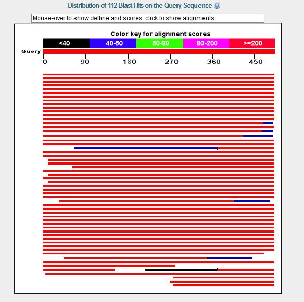
DnaB helicase is not similar from other helicase families. However, they share similarities including triphosphate binding Gly-Lys-(Ser/Thr) motif and the glutamate that binds the "hydrolytic magnesium"
Mechanism of Helicase
To unwind duplex DNA, DnaB helicase reqiures a 5'-overhang single strand DNA; helicase is a 5' to 3' helicase that travels along the lagging strand as it unwinds duplex DNA. Several helicases, which tend to be dimeric, in E. coli travels from 3' to 5' direction along the leading single strand. DnaB helicase subunits are arranged so that there is a hole in the centre into which the single strand DNA fits. Its mechanism is similar to that of F1-ATP synthase. F1-ATP synthase carries out its function using a tight, loose, open mechanism in which ATP is bound to the tight side, ADP and Pi are bound to the lose side and nothing is bound to the open side. A proton gate causes the structure of the sides to shift so that the tight side becomes lose and releases ATP while the lose side becomes tight and forms ATP and the open side becomes lose and binds ADP and Pi. However, in DnaB helicase ATp is hydrolyzed rather than synthesized and the single strand DNA replaces the role of the proton gate. ATP hydrolysis causes the structural shift which result in unidirectional translocation along the single strand DNA.
DnaB helicase consists of 3 distinct domains; domain alpha(R), 1 -156 aa residues; domain beta(â), 157 - 302 aa residues; and domain gamma(ç), 303-471 aa residues. Roles in hexamer formation, DNA-dependent ATPase and DNA helicase activities were examined by using mutants with deletion in the domains. Mutant DnaBâç protein formed a hexamer, whereas mutant DnaBRâ only formed dimers. Dimerization of DnaBRâ is Mg2+ dependent, this shows that oligomerization of DnaB helicase involves at least 2 distinct protein-protein interaction sites. One of these sites its located within domainâ while the other interaction site is located within domainç.
The mutant DnaBâ which is a polypeptide of 147 aa, displayed a completely functional ATPase activity. Therefore, this domain constitutes the "central catalytic domain" for ATPase activity. ATPase activity of DnaBRâ and DnaBâ were kinetically similar, this indicates that domain R had little or no influence on ATPase activity. ATPase activities in both cases were DNA-independent. DnaBâç and wild-type DnaB protein had have comparable DNA-dependent ATPase activity, this indicates a specific role for C-terminal domain ç in enhancement of ATPase activity of domainâ as well as DNA binding. Mutant DnaBâç lacked helicase activity showing that domain R is significant for helicase activity.
Major findings were:
1) Domain â contained a functional ATPase active site.
2) Domain ç is the DNA binding domain and a postive regulator of ATPase activity of domain â.
3) Domain R have no significant effect on ATPase, DNA binding activity or hexamer activity, however, it plays an important role in transducing energy of ATP hydrolysis to unwind DNA by the hexamer.
4) All three domains are required for helicase activity.
Different bacteria has different dnaB helicase protein sequence
Example:
dnaB helicase in E. coli K12
number of aa = 471
       1 magnkpfnkq qaeprerdpq vaglkvpphs ieaeqsvlgg lmldnerwdd vaervvaddf
      61 ytrphrhift emarlqesgs pidlitlaes lerqgqldsv ggfaylaels kntpsaanis
     121 ayadivrera vvremisvan eiaeagfdpq grtsedlldl aesrvfkiae srankdegpk
     181 niadvldatv arieqlfqqp hdgvtgvntg yddlnkktag lqpsdliiva arpsmgkttf
     241 amnlvenaam lqdkpvlifs lempseqimm rslaslsrvd qtkirtgqld dedwarisgt
     301 mgillekrni yiddssgltp tevrsrarri arehggigli midylqlmrv palsdnrtle
     361 iaeisrslka lakelnvpvv alsqlnrsle qradkrpvns dlresgsieq dadlimfiyr
     421 devyhensdl kgiaeiiigk qrngpigtvr ltfngqwsrf dnyagpqydd e
dnaB helicase in H. pylori J99
number of aa = 486
       1 mdhlkhlqql qnierivlsg ivlanhkiee ihsvlepsdf yypphglffe ialklhevnc
      61 pidenfirqk mpkdkqised dlvaifaasp idnieayvee iknasikrkl ftlantireq
     121 alesaqkssd ilnaverevy allngstieg frgikevles tmnlitenqr kgslkvtgip
     181 tgfvqldnyt sgfnqgslvi lgarpsmgkt slmmnmvlsa lnddrgvavf slemsaeqla
     241 lralsdltsi nmhdlesarl dddqwenlak cfdhlsqkkl ffydksyvrm dqirlqlrkl
     301 ksqhkelgia fidylqlmsg nkatkerheq iaeisrelkt lareleipii alvqlnrsle
     361 nrddkrpils dikdsggieq dadivlflyr gyiyqmraed nkidklkkeg kveeaqelhl
     421 kvneerrihk qngsieeaei ivaknrngat gtvytrfnap ftryedmpvd shleegqetk
     481 femptt
dnaB helicase in Geobacillus stearothermophilus
number of aa = 454
       1 mselfserip pqsieaeqav lgavfldpaa lvpaseilip edfyraahqk ifhamlrvad
      61 rgepvdlvtv taelaaseql eeiggvsyls eladavptaa nveyyarive eksvlrrlir
     121 tatsiaqdgy tredeidvll deadrkimev sqrkhsgafk nikdilvqty dniemlhnrd
     181 geitgiptgf teldrmtsgf qrsdliivaa rpsvgktafa lniaqnvatk tnenvaifsl
     241 emsaqqlvmr mlcaegnina qnlrtgkltp edwgkltmam gslsnagiyi ddtpsirvsd
     301 irakcrrlkq esglgmivid ylqliqgsgr skenrqqevs eisrslkala relevpvial
     361 sqlsrsveqr qdkrpmmsdi resgsieqda divaflyrdd yynkdsenkn iieiiiakqr
     421 ngpvgtvqla fikeynkfvn lerrfdeaqi ppga
22 different aa and its abbrev.
Alanine--------------A---------Ala
Cysteine------------C---------Cys
Aspartic acid------D---------Asp
Glutamic acid-----E---------Glu
Phenylalanine-----F---------Phe
Glycine--------------G---------Gly
Histidine------------H---------His
Isoleucine-----------I---------Ile
Lysine---------------K---------Lys
Leucine-------------L---------Leu
Methionine--------M---------Met
Asparagine--------N---------Asn
Pyrrolysine--------O---------Pyl
Proline--------------P---------Pro
Glutamine---------Q---------Gln
Arginine------------R---------Arg
Serine---------------S---------Ser
Threonine----------T---------Thr
Selenocysteine---U---------Sec
Valine---------------V---------Val
Tryptophan--------W--------Trp
Tyrosine------------Y---------Tyr
--ThomasHuber 09:37, 19 February 2008 (EST)
[[Image: [[Image:
Headline text
]]]]