Uploads by Kjafferi
From MDWiki
Jump to navigationJump to search
This special page shows all uploaded files.
| Date | Name | Thumbnail | Size | Description | Versions |
|---|---|---|---|---|---|
| 00:00, 12 June 2007 | Fig6 residues.jpg (file) |  |
33 KB | 4 | |
| 23:20, 11 June 2007 | Fig1 properties.jpg (file) |  |
233 KB | 4 | |
| 04:35, 11 June 2007 | Crystal structure of phosphoserine phosphatase from Methanococcus jannaschii, a hyperthermophile, at 1.8 A resolution.pdf (file) | 855 KB | 1 | ||
| 03:43, 11 June 2007 | Fig1 properties.JPG (file) |  |
176 KB | 1 | |
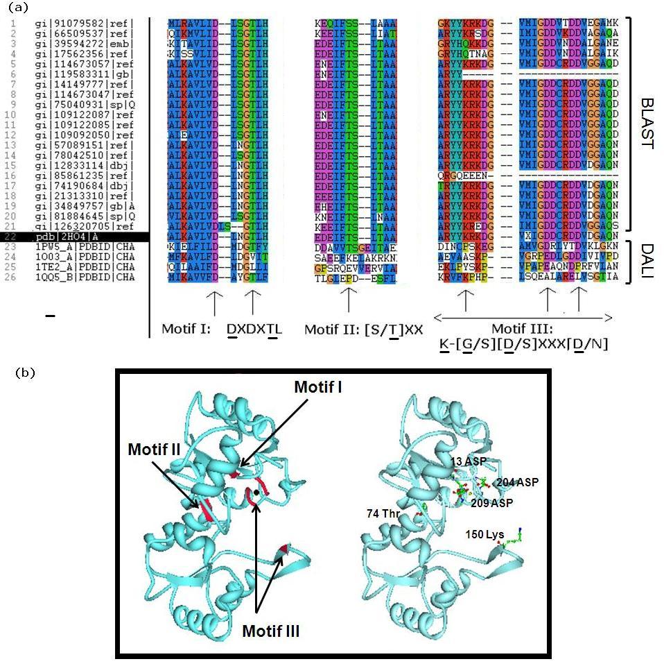
| 03:35, 11 June 2007 | Fig5 MSA.jpg (file) |  |
186 KB | 4 | |
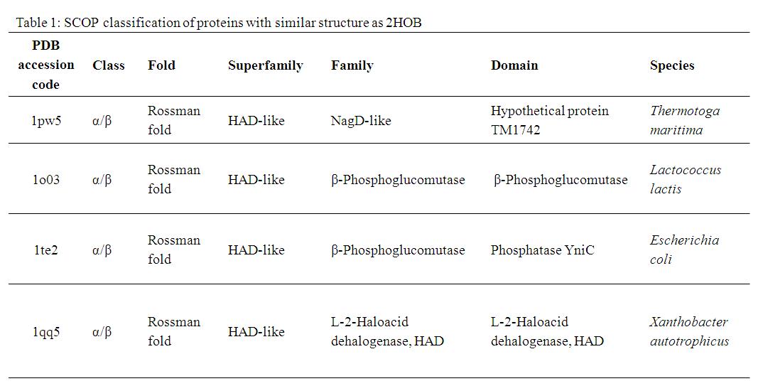
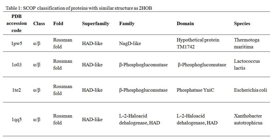
| 03:23, 11 June 2007 | Table1.jpg (file) |  |
68 KB | 3 | |
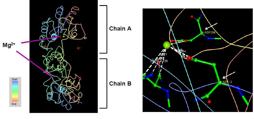
| 10:08, 10 June 2007 | Fig3 Mg.jpg (file) |  |
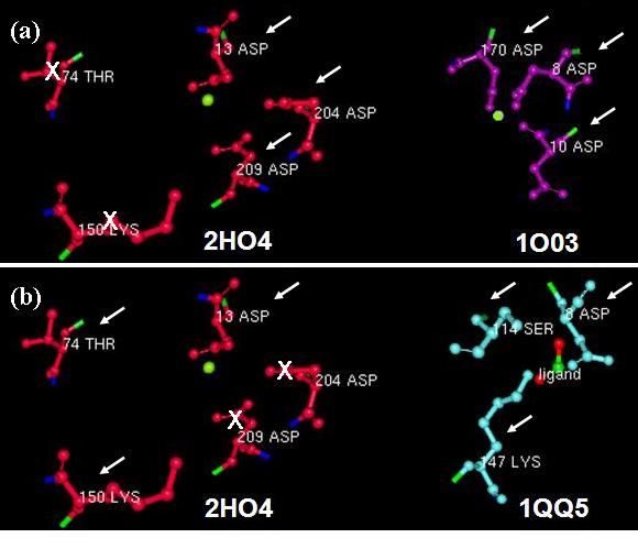
66 KB | Fig 3. The Mg ions are shown in green, sits in 2HO4 interacting with the residues Asp 13 and Asp 204 on chain A. | 3 |
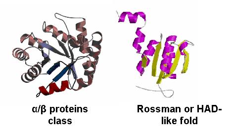
| 09:19, 10 June 2007 | Fig4 class&fold.jpg (file) |  |
21 KB | Fig 4. The α/β protein class and the Rossman fold. These are the most probable classifications for the 2HO4. | 1 |
| 09:13, 10 June 2007 | Table1.bmp (file) |  |
69 KB | 1 | |
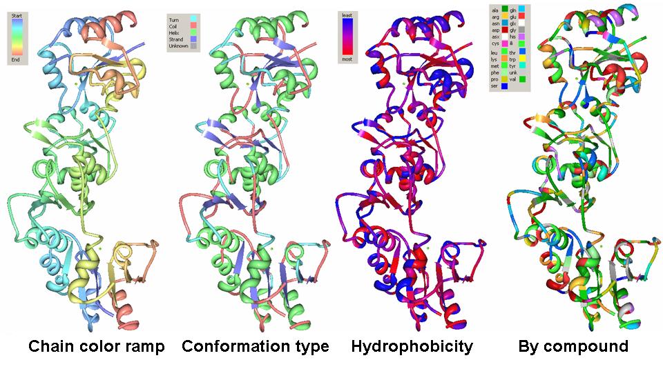
| 08:57, 10 June 2007 | Fig2 properties.jpg (file) |  |
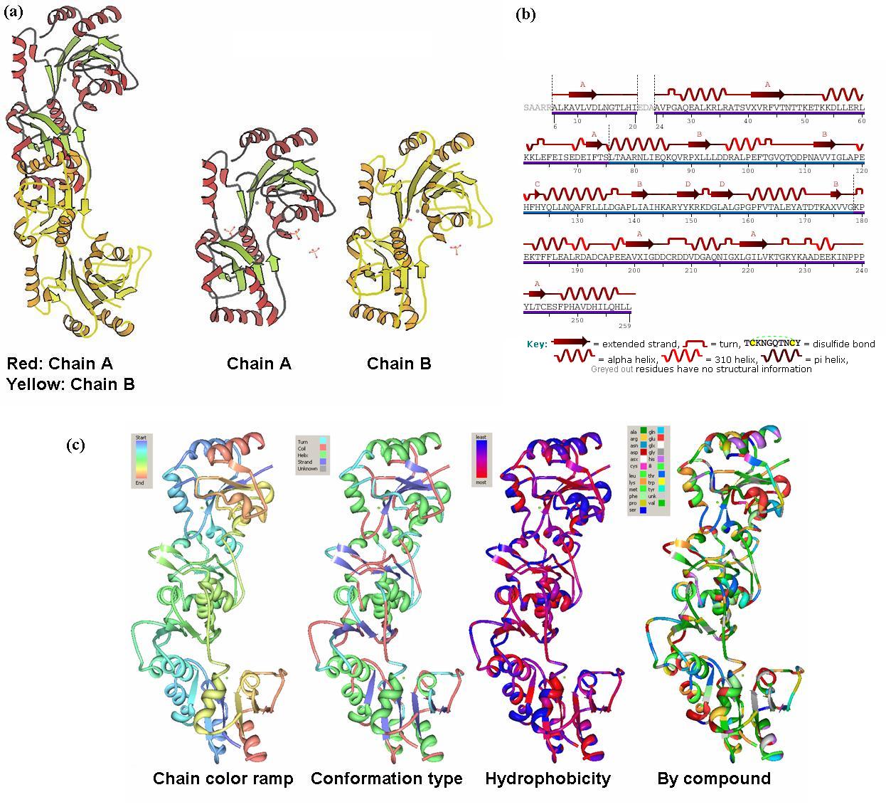
110 KB | Fig 2. The conformation type, hydrophobicity, and the color-coded residues of chain A and chain B in 2HO4. The pictures were generated using the PDB protein workshop. | 1 |
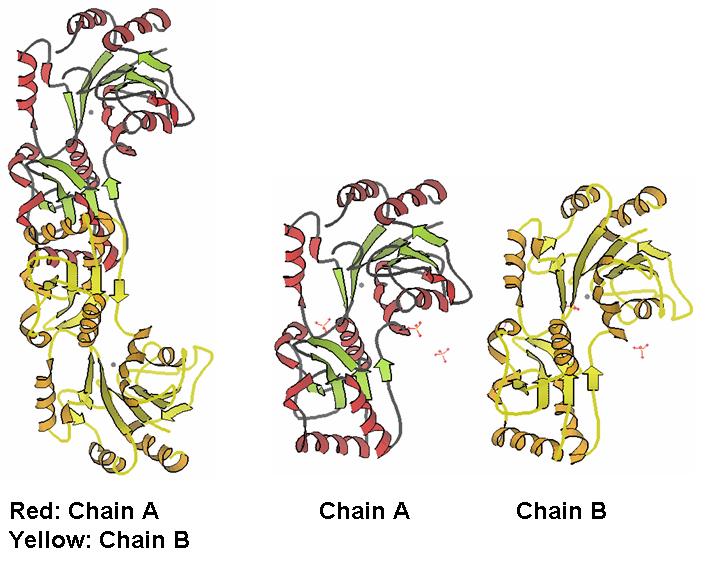
| 08:44, 10 June 2007 | Fig1 chains.jpg (file) |  |
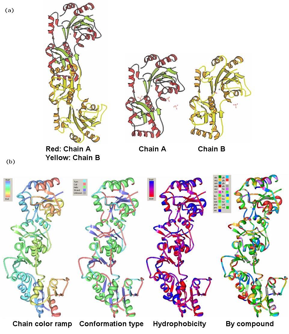
66 KB | Fig 1. The monomers of 2HO4 forming dimmers. Chain A and chain B are exactly similar. | 2 |
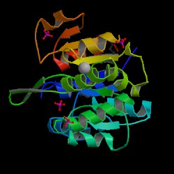
| 06:26, 1 May 2007 | PDB structure.jpg (file) |  |
15 KB | 2 |