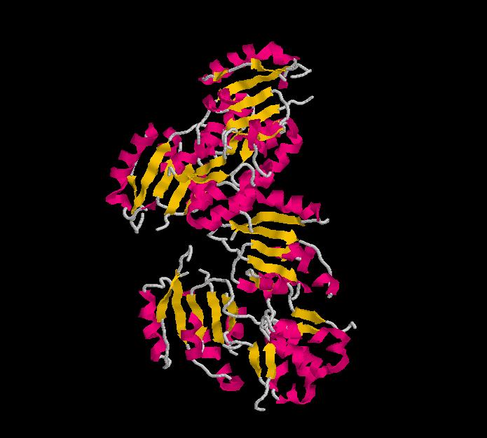
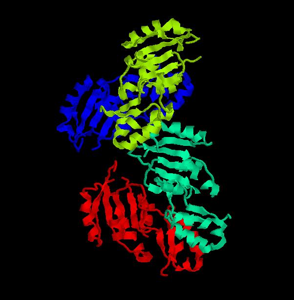
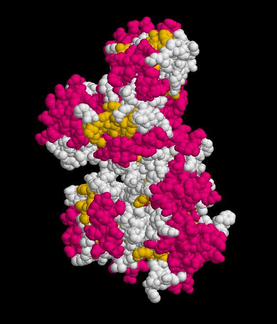
Structure.
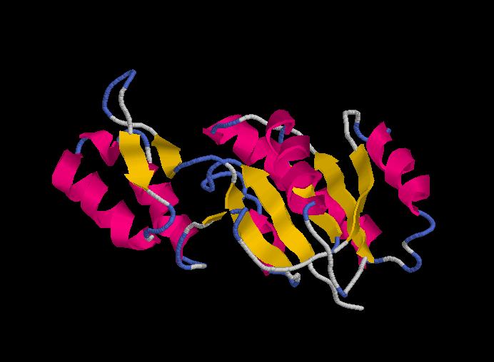
Structure of Ssu72 RNA polymerase II CTD phosphatase homolog
PDB ID: 3fdf
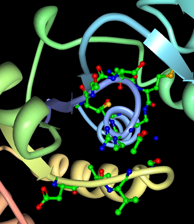
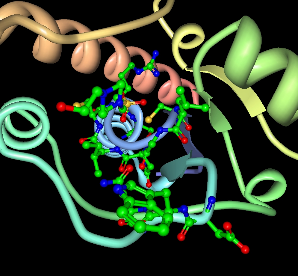
Structure determined using XRD. The following images were obtained using RasMol.
Protein Structure Database Searching by DaliLite v. 3
The Dali server is a network service for comparing protein structures in 3D. You submit the coordinates of a query protein structure and Dali compares them against those in the Protein Data Bank (PDB).
These are the chains of different molecules that best matched to the Ssu72 RNA polymerase II CTD phosphatase homolog (it is important to notice that the first match corresponds to the protein that is being researched).
PDB ID ... Z rmsd lali nres %id PDB Description
3fdf-A..... 37.3 0.0 191 191 100 PDB MOLECULE: FR253;
3fmv-H... 32.4 0.6 190 190 100 PDB MOLECULE: SERINE PHOSPHATASE OF RNA POLYMERASE II CTD;
1dg9-A... 11.4 2.3 120 157 13 PDB MOLECULE: TYROSINE PHOSPHATASE;
1rxi-A...... 9.9 2.4 109 131 15 PDB MOLECULE: ARSENATE REDUCTASE;
DaliLite results: Multiple Structure Alignment
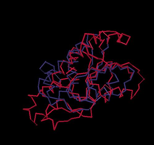
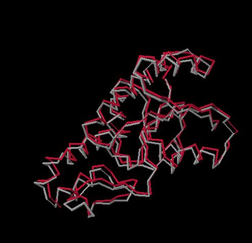
DalaLite results: Superimposed structures
FR253-TYROSINE PHOSPHATASE
FR253-ARSENATE REDUCTASE
FR253-SERINE PHOSPHATASE OF RNA POLYMERASE II CTD
3fdf vs 1dg9
Drosophila
Tyrosine Phosphatase
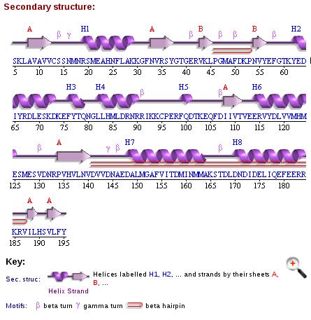
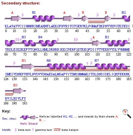
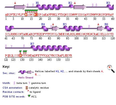
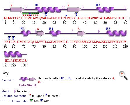
Secondary Structures
FR253
SERINE PHOSPHATASE OF RNA POLYMERASE II CTD
TYROSINE PHOSPHATASE
ARSENATE REDUCTASE
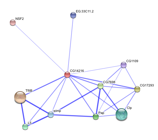
STRING - Known and Predicted Protein-Protein Interactions
Homo sapiens homolog
Structure obtained through Swiss-Model Automatic Modelling Mode
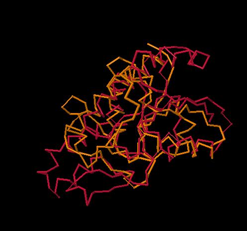
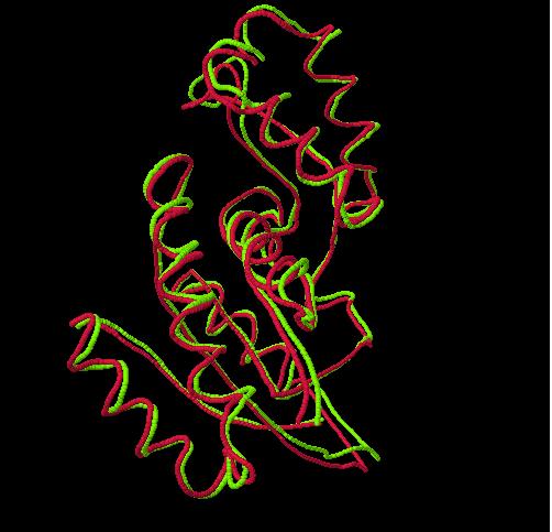
Superimposition, using Dali server, between Homo sapiens homolog and 3fdf-A
Structure Alignment
