Structure for haloacid dehalogenase-like hydrolase domain containing 2
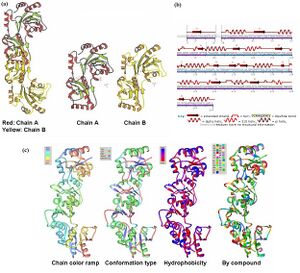
The information on the PDB website suggested that 2HO4 is a hydrolase belonging to the haloacid dehalogenase (HAD) superfamily, consisting of 2 domains. It consists of 259 residues and has a molecular weight of 29 kDa. Moreover, the PDB structure viewers demonstrated that 2HO4 is a monomer but forms a dimer with itself to give Chain A and chain B (Fig 1a).
The PDB workshop enabled us to examine the various properties of 2HO4 including its conformation type and hydrophobocity. Its secondary structure mainly consists of alpha helices and beta sheets with 44% helical (14 helices; 123 residues) 17% beta sheet (12 strands; 48 residues) as shown in Fig 2. The two chains bind near their hydrophobic regions to produce dimeric structures. The outer surfaces of the dimeric protein is hydrophilic whereas the more hydrophobic regions lie near the site of binding (Fig 1b).
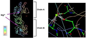
2HO4 has two different ligands i.e a magnesium ion and three phosphate ions (Fig 1). The PDB structures of 2HO4 indicate that the Mg2+ ion binds between the coordinates Asp 13, and Asp 204 on each of the chains as shown in (Fig 2).
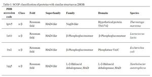
A Dali search with a strict threshold of Z > 12 for 2HO4 found 10 matches. Of these 10 protein matches, we found that most of them were hydrolases belonging to various different families. We reduced our structural matches of 10 proteins to only the ones that had been previously annotated. These proteins had sequence identities within the range of 17% to 25% and they included a putative NagD-like protein of Thermotoga maritima, the β-phosphoglucomutase of Lactococcus lactis , the β-phosphoglucomutase of Escherichia coli, and the L-2-Haloacid dehalogenase of Xanthobacter autotrophicus. The results are summarized in Table 1.
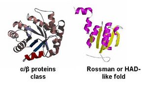
By looking at the architectural classification (using SCOP) of these proteins, it was noted that all of these proteins structures consisted of the same class, fold, and superfamily. The class was alpha and beta (α/β), which contains many parallel beta sheets (beta-alpha-beta units). Likewise, all the proteins had a HAD-like fold consisting of three layers of α/β/α parallel beta-sheet, which amy also be called a Rossman fold (Fig 3). And the superfamily they belonged to was also HAD-like, which usually contains an insertion (sub)domain after strand 1.
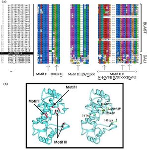
Multiple sequence analysis of H2O4 using the sequence homologs from BLAST as well as the structural homologs from DALI shows that it contains the three highly-conserved sequence motifs of the HAD superfamily, indicating that 2HO4 is a member of this superfamily (Fig 4a). The three motifs are: motif I: DXDX[T/V][L/N]; motif II: [S/T]XX; and motif III: K-[G/S][D/S]XXX[D/N]. These motifs lie next to each other and next the Mg2+ ion, together defining the active site of 2HO4 (Fig 4b) | (Rangarajan et al, 2006).
.
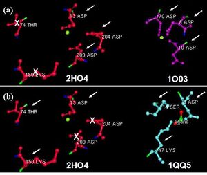
Dali revealed the β-Phosphoglucomutase of Lactococcus lactis (PDB accession code 1O03) to be structurally similar to 2HO4 with 24% sequence identity. We found that they had the three residues in common, which were conserved in the three HAD motifs. Figure 5 shows a comparison of the common residues in 2HO4 and 1O03 present near their active sites (Fig 5). Besides the architectural similarity between 2HO4 and 1O03, 1O03 also consists of a Mg2+ ion bound to its catalytic site. It is possible that the two proties share similar reaction mechanisms.
