COASY discussion
CoA is an essential cofactor in living cells, synthesised from pantothenate (vitamin B5) by a five step pathway that is shared by both prokaryotes and eukaryotes. In eukaryotes, the final two steps of this pathway are catalysed by a single bifunctional enzyme, CoAsy, and previous research has confirmed that mouse CoAsy is a 563 amino acid protein encoded by the Ukr1 gene, and possessing two domains (DPCK and PPAT).
Initial analysis of CoAsy expression confirmed that the expression of CoAsy is consistent with its function. CoA is involved in widespread metabolic processes (eg. the tricarboxylic acid cycle) and the baseline expression of CoAsy in all tissues facilitates synthesis of the CoA needed in for these processes. However, CoA is also involved in other processes such as fatty acid synthesis, which occurs primarily in the liver and adipose tissue (Fox, 2004, p115). This is consistent with the higher levels of CoAsy expression in these tissues. The reason for the high level of CoAsy expression in the kidney is, however, less clear. Also, while baseline expression of the other enzymes involved in CoA synthesis (pantothenate kinase, phosphopantothenoylcysteine synthase, phosphopantothenolycysteine decarboxylase) was also present in all tissues, their expression was otherwise quite dissimilar to that of CoAsy. This may have been due to use of intermediates of CoA synthesis in other tissues, however, this possibility requires further investigation. Nonetheless, the elevated expression of CoAsy in the adipose tissue, and particularly in the liver, is consistent with its function in CoA synthesis. Similarly, the discovery of Zhyvoloup et. al. (2003) that CoAsy is associated with the mitochondrial outer membrane is consistent with its function in CoA synthesis, as CoA is used in the mitochondria during the tricarboxylic acid cycle (Fox, 2004, p108).
The crystal structure of part of CoAsy has recently been determined, and by analysing this structure we have shed further light on the structural composition and mechanism of action of this essential enzyme. Although CoAsy is yet to be formally structurally classified for fold and homology, based on other structurally related proteins it is almost certain to be of the Rossmann A-B-A topology of folds, and to be of the P-loop containing nucleotide triphosphate hydrolases homology Table 2.
Bioinformatic analysis revealed that CoAsy chain A contains the DPCK domain of CoA and part of its PPAT domain. The presence of the DPCK domain was reflected in the high structural similarity of CoAsy chain A to several other kinases, including polynucleotide kinase, adenylate kinase, deoxyguanasine kinase and adenosine-5’phosphosulfate kinase. However, most of the PPAT domain was not present on chain A, as it was positioned before residue 295 of the full-length protein, which was removed during structural determination.
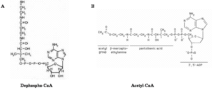
However, the whole of the DPCK domain was present on chain A, and putative binding sites for its ligands (ATP and dephospho-CoA) were determined. Strong evidence suggested that ATP binds in the region of the DPCK domain shown in Figure 6. Pattern searches revealed that a P-loop (an ATP/GTP binding motif) was present in this region. It contained S72 and G75, which were predicted ligand binding residues. Moreover, proteins identified as being structurally similar to CoAsy (chain A) showed particularly strong structural similarity in this region (see Figure 11). The MSA shows that the same was true for sequence similarity (see Figure 14). Together, this strongly suggests that ATP binds in the region shown in Figure 6. In future, this could be confirmed by experimental approaches. Dephospho-CoA may bind to the DPCK domain between the region of its 5th and 7th alpha-helices. During structural determination, acetyl-CoA cocrystallised in this region of CoAsy. Because of the similar structure of dephospho-CoA and acetyl-CoA (see Figure 16), we have predicted that this region may correspond to the dephospho-CoA binding site. This is supported by the presence of predicted ligand binding residues in this region (R128, L131, V135, F136, M142, L145, T146, W150, I153). It is also suggested by the similarity of this region in different species, shown on our MSA (see Figure 14). However, the position of the binding site and the precise residues involved needs to be further confirmed by experimental approaches, ideally using dephospho-CoA rather than acetyl-CoA. This is particularly necessary because predicted ligand binding residues and sequence similarity did exist in regions of the protein that were not predicted to bind ATP or dephospho-CoA.
Because most of the PPAT domain was not present on CoAsy Chain A, binding sites for its ligands (ATP and 4’ phosphopantotheine) could not be determined. These binding sites did not appear to reside on the small part of the PPAT domain that was present, as they were not revealed by pattern searches.
Also, because it did not contain the PPAT domain, chain A could not be used to determine the structural interaction between the PPAT and DPCK domains of CoAsy. Further studies to determine the structure and ligand binding sites of the PPAT domain of CoAsy, together with our predictions of the ligand binding sites on the DPCK domain, could shed valuable light on the interactions between the two domains and the functional significance of these. This may, for example, help to determine whether tunnelling of dephospho-CoA occurs between the PPAT and DPCK domains. This could help resolve the controversy over whether the lack of dephospho-CoA accumulation in cells is due to PPAT being a rate-limiting enzyme, or, as Daugherty et. al. (2002) suggested, to this tunnelling effect. Whilst the domains occur on opposite sides of the protein, the missing string of 282 residues constituting the remainder of the PPAT domain could wrap around the back of the structure and receive dephospho-CoA through the opening on the rear side of the ACO cleft (see Figure 6). Further evidence of this can be seen in Figure 13 where the cleft to which ACO is associated is shown to wrap around to the rear side of the protein. This would then allow the DPCK domain to tunnel its by product dephospho-CoA to the PPAT domain for the next step in the pathway. As the current structurally determined A strand of CoAsy was not found to contain the binding site for dephospho-CoA on the PPAT domain, it is then quite possible that the missing segment has this binding site and is able to receive dephospho-CoA through this mechanism.
While we have focussed on mouse CoAsy, CoA synthesis is a universal pathway, and other species have enzymes similar to mouse CoAsy. In humans, the coasy gene coding for bifunctional CoA synthase is located on chromosome 17q12-21 (Aghajanian & Worrall, 2002). The gene is composed of three domains, two of which have known function. The N-terminal domain ranges from 1-179 amino acids and has unknown function, the amino acids 180-358 compose the central domain that encodes for PPAT, and the C terminal domain of amino acids 359-565 codes for DPCK (Daugherty et al., 2002). Coasy is known to have 10 exons and to produce a protein 565 amino acids in length (Aghajanian & Worrall, 2002). In addition to the DPCK domain found in the bifunctional protein, metazoans have an adjunct monofunctional DPCK domain (Genschel, 2004) which is also found on chromosome 17 (Daugherty et al., 2002).
Studies on bacterial species have shown that the CoA synthase activity is produced by PPAT and DPCK operating as monofunctional proteins (Aghajanian & Worrall, 2002). No bifunctional CoA synthase exists in bacterial species or in any other organisms aside from metazoans (Genschel, 2004). Thus, in bacteria, a separate gene codes for each protein, coaD codes for PPAT and coaE encodes DPCK (Daugherty et al., 2002). Although PPAT and DPCK are functional homologues in bacterial and metazoan species, only the DPCK domain of coasy shares sequence similarity to coaE while the PPAT domain of coasy has no sequence similarity to coaD (Daugherty et al., 2002).
The findings from the multiple sequence alignment support research by Daugherty (2002) that the DPCK but not the PPAT domain shares sequence homology between bacterial and metazoan species. It is clear that the metazoans share closely aligned sequence and structural similarities, while the bacteria and some lower eukaryotes are not closely aligned to the metazoans. This confers with current research, which has shown that bacterial CoA synthase activity is produced by two monomer proteins rather than a single bifunctional protein (Aghajanian & Worrall, 2002). However, it has been found that the metazoans have an additional, bacterial derived DPCK domain as well as the domain in the bifunctional protein (Genschel, 2004).
The conservation of the ATP and ACO binding sites (as seen in Figure 14) in many of the species indicate that these regions are important functional sites for binding. This is supported by structural data in Figure 12 showing that the P-loop in the ATP binding domain is one of the most highly conserved structural elements in the enzyme.
The phylogenetic tree illustrates and adds further evidence to the finding that bacteria share a singular domain with the metazoan species. This is evident from the separate and distant branch types of the bacteria as compared to the metazoans. The species with a bifunctional CoAsy are grouped together on the tree while the bacterial species are on an outlying branch. However, it is uncertain why the mosquitoes, fruit flies and beetles are on a separate branch from the predominant mammalian branch while species such as Synechococcus sp, Leishmania braziliensis and Paramecium tetraurelia are included in the mammalian grouping. Previous phylogenetic studies have shown that Plasmodium falciparum to have a similar pattern of CoA genes to metazoans, which may explain the somewhat strange placing of Paramecium tetraurelia in the tree. It is also possible that Synechococcus, Leishmania and Paramecium have diverged from the bacterial groups to develop a CoAsy bifunctional enzyme. This would be an interesting study in evolution if this proposal is proved to be true.
Abstract | Introduction | Results | Discussion | Conclusion | Method | References
